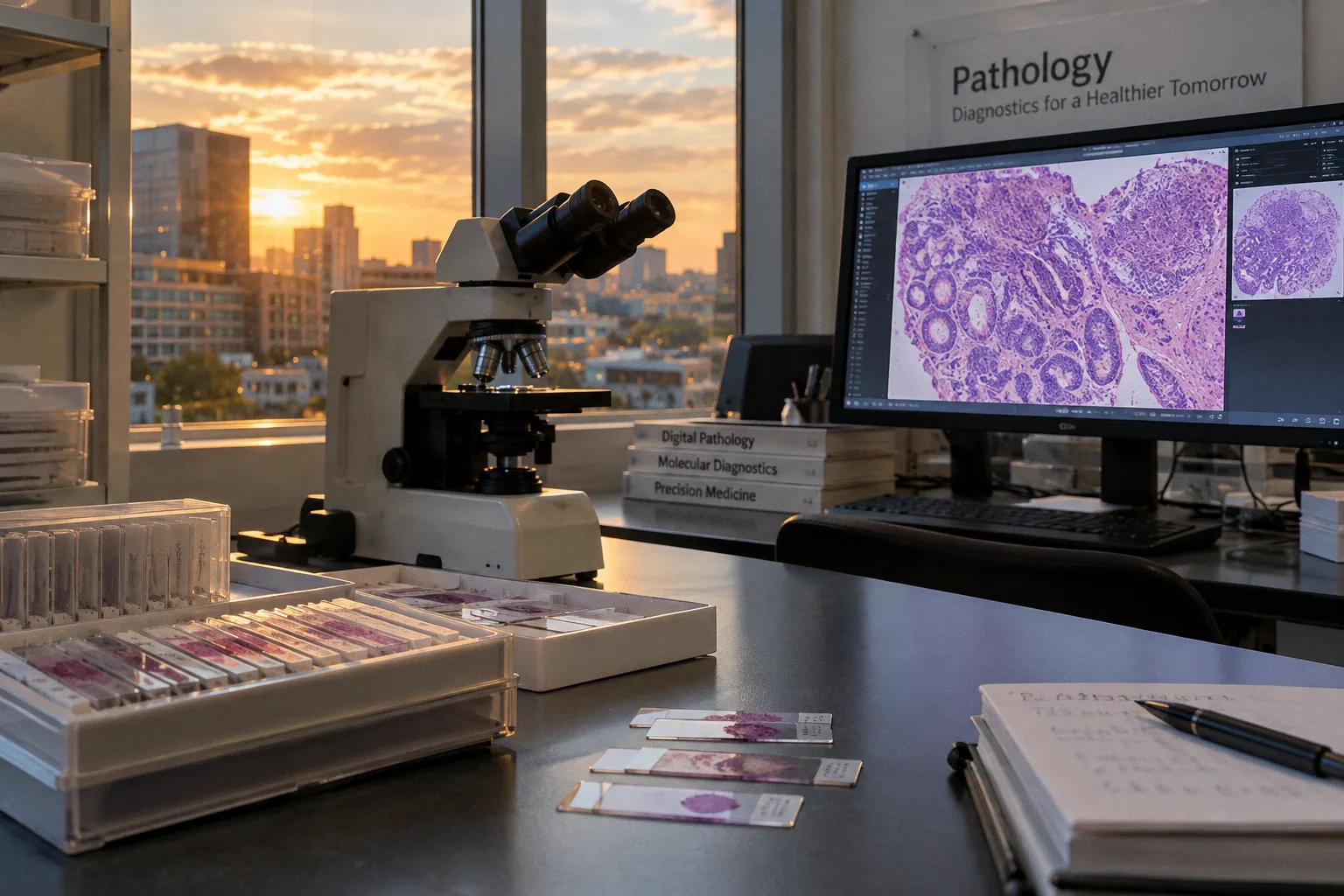
According to healthcare-in-europe.com, Michigan Medicine has become the first US healthcare system to deploy a DICOM-based digital pathology PACS linked directly to its enterprise radiology networks — a structural milestone in cross-departmental image interoperability. The institution subsequently published detailed strategies and infrastructure blueprints documenting how the integration was operationalized across clinical and AI-driven workflows. For pathology laboratories navigating the transition from glass slide microscopy to enterprise-grade digital archives, the deployment provides a reference architecture rather than a marketing proof of concept.
Infrastructure blueprint and workflow integration
The Michigan Medicine deployment demonstrates a practical convergence between two historically siloed imaging domains: whole-slide imaging and DICOM-native radiology systems. By collapsing pathology and radiology onto a shared PACS substrate, the institution has reportedly enabled cross-departmental image retrieval, unified viewer access, and structured handoffs compatible with downstream AI inference pipelines. As reported, the blueprint emphasizes AI interoperability as a foundational requirement — not an overlay — with image metadata, provenance, and annotation schemas engineered for computational consumption from the outset. For laboratory directors, the relevant variable is no longer whether to digitize, but whether the resulting data layer can be ingested by classification, segmentation, and biomarker-quantification algorithms without manual reconciliation.
Parallel signals from spatial analytics and theranostics
The broader landscape is converging around similar architectural assumptions. Voice of Healthcare reports that Lunit and 10x Genomics have entered a collaboration spanning AI pathology and spatial data, suggesting that tissue-level spatial transcriptomics and deep-learning inference are increasingly being treated as complementary analytical strata rather than competing modalities. Separately, Imaging Technology News notes that GE HealthCare's StarGuide GX1 digital 4D CZT SPECT/CT system is now 510(k) pending with the US FDA following its European CE mark, with the platform positioned to advance molecular imaging and precision theranostics workflows, including the acquisition of alpha-emitting isotopes. The connective tissue across these developments is a shared bet on DICOM-native interoperability, structured metadata, and AI-ready data pipelines as the substrate for next-generation precision diagnostics.
What laboratories should track
The Michigan Medicine case offers three operational checkpoints for pathology leaders evaluating analogous deployments: validation of cross-departmental viewer performance under clinical load, demonstrable AI model interoperability against the unified archive, and reconciliation of slide-scanning throughput against radiology image-acquisition SLAs. As the regulatory and commercial ecosystem continues to converge — spanning spatial biology platforms, theranostic imaging systems, and enterprise PACS consolidation — the laboratories positioned to translate these signals into validated, reimbursement-aligned workflows will define the next stratum of molecular diagnostics.