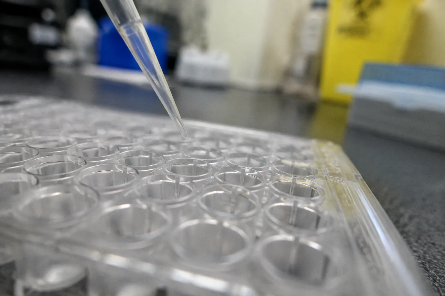
Post-amplification, reaction tubes harbor target amplicons at concentrations ranging from 10¹⁰ to 10¹² copies per microliter—a saturating load of template DNA that renders the surrounding laboratory environment vulnerable to cross-reaction seeding. In accredited diagnostic facilities operating under rigorous contamination controls, carryover rates remain below 1%, yet that residual margin demarcates the boundary between clinically actionable molecular data and the false-positive cascades that compromise patient stratification, antimicrobial stewardship, and syndromic pathogen reporting.
The challenge is structurally embedded in the technology itself. Multiplex assays, which interrogate dozens of pathogen targets within a single reaction vessel, generate not one but a constellation of amplicons, each defined by distinct primer pairs, sequence compositions, and lengths. The very stratification that delivers diagnostic breadth also creates a contamination surface area orders of magnitude larger than that of singleplex PCR. Every opened tube, every ejected pipette tip, every transient breach of containment transfers template into the wider laboratory airspace, where it can persist on surfaces, in reagents, and within instrumentation for extended periods.
The Physics of Contamination: Why Multiplex PCR Is High-Risk
Amplicon carryover contamination is, in mechanistic terms, the unintended introduction of previously amplified DNA products into a freshly prepared reaction, where they serve as spurious templates that generate false-positive signals indistinguishable from bona fide target amplification. The phenomenon is amplified by the extraordinary yield of modern PCR chemistries: a standard 25-microliter reaction can produce billions to trillions of target copies, concentrated into a reaction volume that, once aerosolized, disperses template across benchtops, pipettes, keyboards, and reagent stocks.
The risk profile of multiplex assays is further elevated by the operational reality of high-throughput syndromic panels. A single respiratory pathogen panel may simultaneously amplify sequences from SARS-CoV-2, influenza A/B, RSV, and a dozen additional targets in a closed-tube format, but the pre-analytical steps—nucleic acid extraction, master mix preparation, and sample loading—each introduce discrete opportunities for aerosol-mediated transfer. The consequence is a false-positive signal that may not surface until the analytical phase, when a weak amplification curve on a target that should be absent prompts an investigation that consumes technologist time and clinical bandwidth.
Contamination events are not uniformly distributed across workflow stages. The post-PCR environment, by definition the locus of highest amplicon density, is where the majority of transfer events originate, but secondary contamination of reagents stored in shared cold blocks, or of multichannel pipettes used for both pre- and post-PCR setups, has been documented as a recurring root cause in laboratory audit findings. The variability of contamination patterns underscores why generalized hygiene measures are insufficient: targeted, stage-specific controls are required.
Unidirectional Workflow: Engineering Physical Separation
The primary physical defense against amplicon carryover is the implementation of a strict unidirectional workflow, in which nucleic acid handling is partitioned into discrete zones that mirror the temporal progression of the analytical process. The canonical architecture comprises three zones: a Pre-PCR area dedicated to reagent and master mix preparation, a Sample Preparation area reserved for nucleic acid extraction and template addition, and a Post-PCR area housing thermal cyclers, amplicon detection instruments, and any downstream analysis platforms. Each zone operates with dedicated equipment—including pipettors, vortex mixers, centrifuges, and laboratory coats—and ideally maintains a clean-to-dirty pressure gradient, with the Pre-PCR area at the highest relative pressure and the Post-PCR area at the lowest, so that any aerosolized template is drawn downstream toward the post-PCR environment rather than back toward reagent preparation spaces.
The efficacy of physical separation depends on rigorous adherence to a one-way movement principle: materials and personnel flow from Pre-PCR through Sample Prep and into Post-PCR, but never in reverse. Returning a pipette from the Post-PCR bench to the Pre-PCR bench effectively transports template into the cleanest zone of the laboratory, where it can seed an entire batch of negative controls. HEPA-filtered ventilation, while standard in modern molecular diagnostics suites, is treated as a supplementary rather than primary control; air exchanges dilute but do not eliminate contamination risk, and the absence of pressure monitoring can mask architectural deficiencies that only surface as contamination events downstream.
Dedicated personal protective equipment is non-negotiable. Laboratory coats designated for the Post-PCR area must not enter the Pre-PCR space, and glove changes are required at every transition between zones. In practice, the workflow discipline that physical separation demands is procedural rather than technological—rooted in training, signage, and supervision—which makes it vulnerable to lapses during high-volume periods when staffing pressure compromises protocol adherence. Laboratories that have conducted internal audits of compliance consistently find that the failure mode is not architectural but behavioral: the zone boundary exists, but a technologist crosses it without changing gloves, or a shared reagent is carried between areas during a staffing crunch.
Physical separation is not a single safeguard but a layered architecture, in which dedicated equipment, dedicated PPE, dedicated bench space, and unidirectional traffic collectively reduce the probability that any single transfer event becomes a contamination event.
Enzymatic Defense: The dUTP/UNG Mechanism Explained
Enzymatic carryover prevention addresses the residual risk that persists even within rigorously separated workflows. The standard implementation involves substituting deoxyuridine triphosphate (dUTP) for deoxythymidine triphosphate (dTTP) in the PCR master mix, producing amplicons that incorporate uracil bases in place of thymine. Before each new reaction is subjected to thermal cycling, Uracil-N-Glycosylase—commonly abbreviated UNG or UDG—is added and the mixture is held at approximately 50°C for two minutes. During this incubation, UNG selectively excises uracil residues from any contaminating amplicons carried over from previous runs, generating apyrimidinic sites that destabilize the DNA backbone. The subsequent high-temperature denaturation step, typically conducted at 95°C, performs two functions simultaneously: it inactivates the UNG enzyme, preventing degradation of the newly synthesized target amplicons, and it fragments the apyrimidinic-containing contaminating templates, rendering them unsuitable as amplification substrates.
The selectivity of the system rests on a critical biochemical distinction: native genomic DNA and plasmid templates, which contain thymidine rather than uracil, are unaffected by UNG activity, while only the uracil-incorporated amplicons from previous reactions are cleaved. This discrimination allows the assay to destroy carryover contamination without compromising the analytical sensitivity for the intended target. The protocol thus represents one of the more elegant applications of base-specific enzymology to diagnostic workflow design.
The limitations of the approach are equally precise. UNG protects exclusively against amplicon contamination from previous runs—specifically, those amplicons synthesized in the presence of dUTP. Native genomic contamination, plasmid contamination introduced through cloning or vector work, and amplicons generated in legacy dTTP-based assays remain invisible to the enzymatic control. Laboratories transitioning from dTTP to dUTP chemistry must therefore validate that prior contamination does not pre-exist in the workflow, and they must apply the dUTP/UNG system across all assays sharing a multiplex panel to avoid cross-protection gaps. Additionally, certain modified polymerases and buffer formulations can exhibit partial resistance to UNG, and incomplete heat inactivation at the denaturation step can permit residual glycosylase activity to degrade the newly synthesized product—a failure mode that manifests as reduced analytical sensitivity rather than false positivity.
| Control Layer | Mechanism | Protects Against | Key Limitation |
|---|---|---|---|
| Unidirectional workflow | Spatial and procedural zone separation | Aerosolized amplicon transfer between zones | Dependent on human adherence; does not eliminate intra-zone transfer |
| dUTP/UNG enzymatic control | Selective cleavage of uracil-containing amplicons | Carryover from prior dUTP-based reactions | Ineffective against native DNA, plasmid, or dTTP-based amplicon contamination |
| Chemical decontamination | Sodium hypochlorite oxidation of nucleic acids | Surface-bound template | Does not penetrate instrument internals; requires subsequent rinse |
| UV cross-linking | Thymidine dimer formation on nucleic acid films | Surface-bound amplicons | Ineffective on liquid solutions; penetration depth limited |
Surface Decontamination Protocols and UV Limitations
Surface decontamination completes the contamination control triad, addressing the residual template that accumulates on benchtops, instrument surfaces, and equipment exteriors despite workflow discipline. The standard regimen combines chemical oxidation with ultraviolet cross-linking. A 10% sodium hypochlorite solution—the widely adopted protocol concentration in molecular diagnostics laboratories—applied to surfaces and allowed a contact time of several minutes, oxidizes nucleic acid backbones and renders amplicons non-amplifiable; subsequent rinsing with DNA-free water or ethanol removes the oxidizing agent, which would otherwise inhibit downstream enzymatic reactions. The procedure is straightforward but exacting: at standard working concentration, bleach delivers reliable nucleic acid degradation while remaining compatible with routine surface materials, though prolonged or repeated contact can still corrode stainless steel and degrade polymer components on instrumentation, and any decontamination protocol must account for material compatibility constraints specific to the laboratory's equipment inventory.
Ultraviolet irradiation, typically delivered by germicidal lamps mounted in biosafety cabinets or dedicated cross-linkers, operates through a different mechanism. UV-C radiation induces thymidine dimer formation and adjacent cross-links in nucleic acid molecules exposed on a surface, preventing their function as amplification templates. The technique is particularly effective when applied to work surfaces, the interior of biosafety cabinets, and the exterior of pipette shafts—locations where amplicon-containing aerosols settle as thin films. Standard exposure protocols, ranging from 10 to 30 minutes of UV irradiation, can reduce surface-bound amplicon titers by several orders of magnitude.
The boundaries of UV decontamination, however, are substantive. UV light cannot penetrate liquid volumes to any meaningful degree, and the efficacy of cross-linking falls sharply as a function of surface topography: shadowed regions beneath equipment, the interior cavities of pipette barrels, and the seams between bench surfaces receive negligible exposure. Furthermore, UV treatment does not distinguish between amplicon and native DNA, and prolonged exposure can degrade reagents stored in transparent containers. The technology functions, therefore, as a targeted adjunct to chemical decontamination rather than a stand-alone sterilant, and laboratory protocols that rely on UV alone routinely demonstrate elevated contamination rates relative to those that integrate all three control layers.
Diagnostic Integrity: Balancing Sensitivity and False Positives
The clinical consequence of amplicon carryover is rarely a single erroneous result; it is more often a pattern of contamination that erodes the diagnostic integrity of an entire panel. A persistent low-level signal on a target that should be absent—whether in negative controls, no-template controls, or patient samples from low-prevalence populations—forces the laboratory into one of two defensive postures. The first is to re-run affected samples, consuming reagents, technologist time, and turnaround capacity. The second is to suppress the analytical signal through stricter thresholding, which paradoxically reduces the assay's analytical sensitivity for low-copy genuine positives and compromises the very diagnostic efficacy that the multiplex panel was designed to deliver.
The trade-off between analytical sensitivity and false-positive risk defines the operational envelope of every high-complexity molecular assay. Syndromic respiratory panels, blood culture identification panels, and gastrointestinal pathogen panels each operate at the limit of what current chemistries can resolve, and the cost of contamination is measured not in reagent waste but in clinical misdirection. A false-positive detection of methicillin-resistant Staphylococcus aureus in a blood culture identification panel can trigger days of unnecessary vancomycin therapy, with attendant nephrotoxicity, cost, and antimicrobial resistance pressure. A false-positive SARS-CoV-2 result in a low-prevalence setting can initiate quarantine cascades, contact tracing operations, and clinical decisions whose downstream effects compound rapidly.
The role of contamination control is to compress the false-positive probability toward zero without compressing the limit of detection upward. Each control layer—physical separation, enzymatic deactivation, chemical and UV decontamination—contributes independently to that compression, and the integration of all three layers is what allows well-managed laboratories to maintain carryover rates below the 1% benchmark. Deviations from the integrated model correlate strongly with contamination events, and laboratories that relax any single layer invariably experience an increase in confirmatory re-runs and clinical consults.
The cost of amplicon carryover is measured not in reagent waste but in clinical misdirection: a single false-positive at the wrong target can redirect therapy, skew epidemiological surveillance, and erode confidence in molecular diagnostics.
Containment as a Diagnostic Paradigm
The architecture of contamination control in molecular diagnostics has matured into a self-contained discipline, one in which workflow design, enzymatic chemistry, and decontamination engineering operate in concert to preserve analytical specificity. The 10¹⁰ to 10¹² copies per microliter post-amplification yield that makes PCR so analytically powerful is the same yield that makes it so vulnerable, and the laboratory's response has been to construct layered defenses that anticipate transfer events before they occur. The dUTP/UNG system, in particular, exemplifies how a single biochemical substitution can be leveraged into an enzymatic fail-safe that operates invisibly within every thermal cycling protocol.
The forward trajectory is defined less by novel chemistries than by the discipline of integration. Closed-system cartridges, point-of-care platforms with sealed fluidic paths, and laboratory information systems that flag anomalous signal patterns in real time are incrementally reducing the window in which contamination can propagate undetected. As syndromic panels expand to interrogate dozens—or eventually hundreds—of targets simultaneously, the contamination surface area grows in proportion, and the marginal value of each additional control layer rises correspondingly. Laboratories that treat contamination control as an architectural commitment rather than a procedural checklist will define the operational standard against which the next generation of molecular diagnostics is validated.