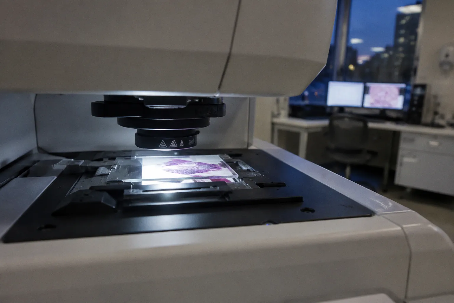
A whole-slide image contains on the order of 10⁹ to 10¹⁰ pixels. Gigapixel territory. No current GPU — consumer or enterprise — can load that at full resolution into VRAM in a single pass. So when someone tells you their model "analyzes whole-slide images," what they actually mean is a chain of preprocessing steps, patch extraction, and aggregation — stitched together into a pipeline that does a lot of heavy lifting before the model even sees tissue.
Let's stress-test that pipeline. Not the marketing version. The engineering one.
The Tiling Problem: Why Patches Are Non-Negotiable
The first constraint is hardware, and it's absolute. GPUs have finite memory — typically 16 to 80 GB of VRAM depending on the card. A single WSI at 40× magnification can occupy several gigabytes of uncompressed pixel data. You're not fitting that into memory, running backpropagation on it, and expecting real-time inference. This is a physics problem, not an algorithm one.
The industry solution is patch extraction. The pipeline divides the slide into smaller sub-images — tiles or patches — commonly sized at 256×256 or 512×512 pixels. Each patch becomes an independent unit of analysis. The model processes thousands of these per slide — sometimes tens of thousands — then aggregates results back to a slide-level prediction.
This is where the "whole-slide" part of WSI analysis quietly becomes a misnomer. You're not analyzing the whole slide in any meaningful architectural sense. You're analyzing local regions and reassembling them. The sophistication lies in how you perform the reassembly.
But before you can extract a single patch, you need to know where the tissue actually is.
Preprocessing: Separating Tissue from Scanner Noise
Not every pixel on a digitized slide contains diagnostic information. Far from it. A typical WSI is mostly background — glass, mounting medium, air bubbles. Pen markings from the pathologist who grossed the specimen. Dust particles from the scanner bed. Streak artifacts from the scanning process itself.
A preprocessing pipeline starts with tissue detection: segmenting the biological material from everything else. This is a binarization step — a mask that says "this region has tissue, this doesn't." It sounds trivial. It isn't. Miss the tissue boundaries and you feed background noise into your feature extractor. Include artifacts and you train your model to recognize pen ink as a morphological feature.
Once the tissue mask exists, the pipeline extracts patches only from regions that passed the filter. Artifacts — pen marks, dust, scanner streaks — get flagged and discarded. The result is a curated set of patches that actually contain analyzable tissue.
This preprocessing stage is pure workflow engineering. No glamour. No ten-thousand-citation papers. But it's the foundation everything else rests on. Garbage patches in, garbage predictions out. Every time.
Stain Normalization: The Color Problem Nobody Solves Cleanly
Here's a friction point that vendors consistently underplay: the same tissue stained with hematoxylin and eosin will look different depending on which lab processed it, which reagent batch was used, and which scanner digitized it. Different scanner optics. Different color temperatures. Different CCD sensitivities. The result is batch effect — systematic color variation that has nothing to do with biology and everything to do with equipment.
For a pathologist, this is manageable. They recalibrate by eye. They adjust to the slide. For a deep learning model, it's a landmine. If your training data comes from one hospital and your deployment data comes from another, the model may fail — not because the disease changed, but because the stain looks different.
The standard countermeasure is stain normalization. The Macenko method, published in 2009, remains widely used across pipelines: it extracts the principal stain vectors from each image and maps them to a reference template. The goal is visual consistency across slides from different labs and scanners.
But here's the reality check: stain normalization mitigates batch effects. It does not eliminate them. No normalization algorithm fully accounts for all the variability across every scanner, reagent, and tissue preparation protocol. It reduces the gap. You still need cross-site validation.
Stain normalization reduces visual noise across labs — it doesn't remove the need for cross-site validation. Treating it as a silver bullet is how models quietly fail in deployment.
Multiple Instance Learning: Scaling Without Manual Labels
The annotation bottleneck is where traditional supervised learning hits a wall in pathology. Pixel-level annotation of tissue on WSIs is expensive, slow, and requires specialist pathologists. You're asking a highly trained clinician to spend hours per slide drawing boundaries around regions of interest. That doesn't scale. Not in any lab I've seen.
Multiple Instance Learning offers a workaround. Instead of requiring pathologists to annotate every patch, MIL trains on slide-level labels. The slide is "cancer" or "not cancer." The model figures out which patches matter.
Architecturally, the slide becomes a "bag" of patches. Each patch is an "instance." The model learns an aggregation function — typically attention-based — that weights patches by their contribution to the slide-level prediction. The CLAM framework, introduced in 2021, popularized attention-based MIL for histopathology and became a go-to architecture for WSI classification.
This is a genuine architectural shift. It moves the annotation burden from "annotate every cell" to "label the slide." For clinical deployment at any meaningful scale, that's the difference between a research demo and a viable pipeline.
But MIL isn't free. The attention mechanism is a black box. Which patches the model considers important — and why — isn't always interpretable. For regulatory submission or clinical trust, that interpretability gap is a problem you need to address separately. It's solvable, but it takes work that most publications gloss over.
Foundation Models and the Pre-Training Scale Shift
The latest evolution in the pipeline is foundation models — large architectures pre-trained on massive datasets of pathology patches before being fine-tuned on specific diagnostic tasks.
Prov-GigaPath, introduced in 2024, trained on approximately 1.3 billion patches. That scale of pre-training produces a general-purpose feature extractor that can be adapted to downstream classification tasks with relatively little labeled data. The idea is transfer learning from a massive, diverse corpus of tissue morphology.
This is where the patch-based pipeline proves its worth beyond being a hardware workaround. Because patches are the atomic unit of analysis, you can pre-train on billions of them across organ types, staining protocols, and institutions. The patch abstraction becomes a universal interface between raw pixels and model architecture.
The trade-off is compute. Pre-training at this scale requires enormous GPU clusters and training budgets that most institutions simply don't have. For the majority of hospital labs and even reference labs, you're not pre-training a foundation model from scratch — you're fine-tuning one. The deployment question becomes practical and specific: which pre-trained model best fits your tissue types, your staining protocols, your diagnostic tasks?
| Pipeline Stage | Problem It Solves | Maturity Level |
|---|---|---|
| Patch extraction | GPU memory constraint at gigapixel scale | Solved; standardized at 256–512 px |
| Tissue detection | Background and artifact filtering | Mature; AtlasPatch achieves >0.98 precision |
| Stain normalization | Cross-site color variation | Mitigated but not eliminated; Macenko still standard |
| Feature encoding | Morphological representation of tissue | Rapidly evolving; foundation models entering clinical research |
| Slide-level aggregation | Bridging patch features to diagnosis | MIL and transformer-based; CLAM widely adopted |
Throughput: The Bottleneck After the Model
The most overlooked part of the pipeline isn't accuracy — it's speed. A single WSI can yield tens of thousands of patches. Across a lab processing hundreds of slides per day, the preprocessing pipeline — tissue detection, patch extraction, artifact filtering, stain normalization — becomes a throughput bottleneck that determines whether your AI model ever actually runs.
AtlasPatch, released in 2026, targets this directly. It achieves a tissue detection precision of 0.986 while delivering up to a 16× speedup in end-to-end WSI preprocessing compared to standard deep-learning pipelines. That's not an incremental improvement. That's a repositioning of where the computational time actually goes.
This matters because the deployment friction for computational pathology AI isn't just "does the model work?" It's "can the model work at lab throughput without adding 45 minutes of preprocessing per slide?" If your preprocessing pipeline can't keep pace with scanner output, the AI sits idle between runs. Workflow integration — the term nobody puts in press releases — is where real-world viability is won or lost.
The AI model gets the headlines. The preprocessing pipeline determines whether it ever runs in production.
Where This Actually Stands
Let's be direct about the maturity picture.
The patch-based pipeline architecture for deep learning in computational pathology is engineering-proven. Tissue detection, patch extraction, stain normalization, MIL-based aggregation — these components work, individually and in sequence. The academic literature has validated them across organ systems and diagnostic tasks. The individual bricks are solid.
What hasn't happened is broad clinical deployment at scale. The friction points remain real. Stain normalization still can't fully bridge scanner-to-scanner variation. MIL architectures lack the interpretability regulators expect from diagnostic tools. Foundation models are compute-heavy, and institution-specific fine-tuning is non-trivial to operationalize. Preprocessing throughput, while improving, needs to consistently match real lab volumes across every deployment.
If you're evaluating a computational pathology AI product today, don't ask about model accuracy in isolation. Ask about the preprocessing pipeline. Ask how it handles stain variation from your specific scanner. Ask what happens at your slide volume, on your hardware, with your tissue types. The model is the headline. The pipeline is the product.
The patch-based approach isn't going anywhere. It's a hardware constraint that became an architectural standard. The question was never whether to use patches — it's whether the pipeline around them is mature enough for your lab's workflow.
Right now, for research and controlled validation studies, it is. For high-volume clinical deployment across multi-site networks? We're close. But the gap between "works in the paper" and "runs in production" is exactly where the next wave of engineering has to happen — and where most of the real work still remains.