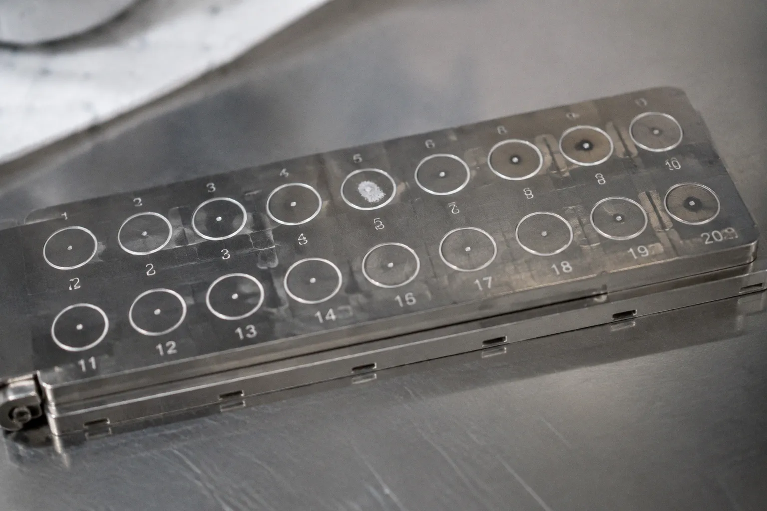
In one diagnostic scenario, the culture showed a non-lactose-fermenting Gram-negative rod. The Gram stain separately demonstrated Gram-negative bacilli, with morphology that supported a non-fermenting enteric isolate but could not, by itself, establish lactose fermentation. MALDI-TOF nevertheless returned Escherichia coli with a confidence score high enough to invite immediate acceptance. That disconnect—between the observable phenotype and the instrument’s confident output—is where real diagnostic risk begins.
Matrix-assisted laser desorption/ionization time-of-flight mass spectrometry has transformed clinical microbiology. A colony is placed on a target plate, treated with matrix, loaded into the instrument, and converted into a protein spectrum within minutes. The laboratory no longer has to wait for a full biochemical panel before obtaining a preliminary identification. The technology has changed the tempo and economics of routine diagnostics.
But speed can quietly erode critical thinking. MALDI-TOF is not reading the organism’s genome in its entirety. It is comparing a protein fingerprint with a reference library. When the spectrum is incomplete, the preparation is poor, or two taxa are proteomically very similar, the instrument may produce a plausible answer that is not the correct one. The most dangerous errors are not always the visibly weak results. They are the wrong identifications accompanied by confidence scores that appear reassuring.
The hard truth is that a concordance rate of 86.8% between MALDI-TOF and conventional identification methods sounds comfortable until the remaining 13.2% is translated into clinical practice. That figure comes from a comparative evaluation of 204 clinical isolates. A discrepancy is not merely a technical disagreement in a spreadsheet. It may change isolation precautions, antimicrobial selection, public health reporting, or the interpretation of a positive blood culture.
The Limits of Spectral Resolution in Closely Related Taxa
MALDI-TOF identifies microorganisms by generating a mass spectrum dominated by abundant proteins, particularly ribosomal and other conserved cellular proteins. The spectrum is then compared with entries in a database. The elegance of the method is also its central limitation: when two organisms share enough of their proteomic profile, the instrument may not have sufficient discriminatory information to separate them reliably.
The Escherichia coli–Shigella boundary is a classic example. These taxa are closely related genetically and phenotypically, and their ribosomal protein spectra overlap substantially. Depending on the platform and database, a routine run may report E. coli for an isolate that is actually Shigella flexneri, or return a Shigella identification for an isolate that belongs to E. coli.
That distinction is not academic. A Shigella identification can trigger public health notification, additional epidemiological investigation, and specific infection-control considerations. An E. coli result may instead be interpreted as an ordinary urinary or enteric isolate. The same colony can therefore lead to very different downstream actions depending on which side of the taxonomic boundary the instrument places it.
Research using custom biomarker models has shown that the complex can be approached more effectively than it is by many routine commercial databases. Reported custom approaches have achieved roughly 90% species-level identification accuracy, with misidentification rates reduced to around 3%. Those models, however, are not necessarily available in the routine workflow of a community hospital laboratory. Most technologists work with the database supplied for their instrument and software version. If that library does not contain sufficiently discriminating entries, a high score cannot manufacture information that the spectrum does not contain.
The same problem appears elsewhere in clinical microbiology. Closely related taxa may produce spectra that are difficult to separate even when their clinical implications differ. The comparative evaluation of 204 isolates identified substantial genus-level discrepancies involving pairings such as:
- Enterobacter and Raoultella, which may differ in clinically relevant resistance expectations and interpretation;
- Streptococcus and Gemella, where morphology and clinical context can be important to the final call;
- Pseudomonas and Burkholderia, where species-level distinctions may affect treatment and infection-control decisions.
The practical point is simple but easy to lose in an automated workflow: a confidence score expresses how well the submitted spectrum matches the database entry. It does not certify that the database contains the right taxonomic distinction, that the colony was pure, or that the result agrees with the patient’s specimen and clinical picture.
MALDI-TOF gives you speed. But when two organisms share nearly the same protein fingerprint, that speed can carry a wrong answer to the chart before anyone has time to question it.
Analyzing the 13.2% Discrepancy Rate in Routine Clinical Isolates
A discrepancy rate measured under study conditions does not behave like a discrepancy rate in a busy laboratory. In a controlled evaluation, isolates are selected carefully, colonies are usually fresh, and the process is observed closely. Routine work is less forgiving. Plates arrive in batches, specimens vary in quality, and a technologist may be moving between Gram stains, culture workups, antimicrobial susceptibility testing, and critical-value calls.
The error is often cumulative rather than dramatic. Each step may appear acceptable in isolation, but several small weaknesses can converge on one unreliable identification.
| Factor | What can go wrong | Why the result may still look convincing |
|---|---|---|
| Database composition | The reference library may contain limited or poorly discriminated entries for a taxon pair | The best available match can receive a strong score even when the correct species is absent or indistinguishable |
| Colony age | Protein expression changes as cultures move beyond active growth | The spectrum may remain readable while losing the features that support accurate discrimination |
| Colony selection | Mixed growth, satellite colonies, or a neighboring morphotype may be picked | The instrument identifies the material placed on the target, not necessarily the organism the technologist intended to select |
| Material on the spot | Too little biomass produces a weak spectrum; too much may contribute to ion suppression or background | A partial spectrum can still generate a reportable match |
| Matrix application | Uneven crystallization or poor mixing can reduce spectral quality | The software may analyze a noisy spectrum without making the source of the noise obvious |
| Calibration | Mass accuracy can drift when calibration is missed or poorly performed | Small shifts are not always visible in the final confidence display |
| Preparation method | A direct smear may be inadequate for organisms with resistant cell envelopes | The result may be low-quality, ambiguous, or falsely matched rather than simply rejected |
| Purity and culture conditions | Subculture conditions can alter expression patterns or preserve contaminants | The database comparison cannot correct for a biologically unsuitable input |
This is why “the machine gave a high score” is not a sufficient final argument. The score is conditional on the quality of the submitted material, the reference library, and the algorithm’s ability to distinguish the available candidates.
The original diagnostic case illustrates the point. The culture showed a non-lactose-fermenting Gram-negative rod. That observation came from growth behavior on appropriate differential media, not from the Gram stain. The Gram stain provided a separate piece of information: the organism was a Gram-negative bacillus. If the culture phenotype and the MALDI-TOF identification do not agree, the discrepancy should be treated as a reason to pause, review purity, repeat preparation, and assess whether the organism belongs to a known blind spot of the database.
A routine laboratory does not need to distrust every MALDI-TOF result. It does need to know which results deserve resistance. A species call is more vulnerable when:
1. The result conflicts with the Gram stain or colony morphology.
2. The organism comes from a normally sterile site.
3. The reported species is unusual for the specimen or patient population.
4. The identification sits within a taxonomic group known to produce overlapping spectra.
5. The confidence score is acceptable but the replicate spots are inconsistent.
6. The isolate has an antimicrobial susceptibility pattern that does not fit the identification.
7. The result would change infection-control, public health, or outbreak-management decisions.
Those are not abstract safeguards. They are ways of preventing an automated identification from becoming an unexamined clinical fact.
Diagnostic Challenges with Streptococcus pneumoniae and the Mitis Group
The distinction between Streptococcus pneumoniae and members of the Streptococcus mitis group is one of the clearest demonstrations of why MALDI-TOF results need biological context.
Streptococcus pneumoniae is associated with pneumonia, bacteremia, meningitis, and other invasive infections. Mitis group streptococci are common upper-respiratory commensals and can also cause genuine disease, but they are frequently encountered as possible contaminants in blood cultures. The identification therefore affects more than the label on a report. It can influence whether a positive blood culture is treated as a serious invasive pneumococcal infection or interpreted as contamination or less clinically significant bacteremia.
The first difficulty is spectral similarity. The protein fingerprints of S. pneumoniae and organisms such as S. mitis and S. oralis overlap sufficiently that routine databases may return an ambiguous result or assign the wrong species. Candidate mass-to-charge markers, including peaks reported around 4,964.32, 6,888.90, and 9,516.46 m/z, have been investigated as potential discriminators. But a useful biomarker is not the same thing as a universally reliable routine rule. Its value depends on the instrument, database, spectrum quality, and the algorithm used to interpret it.
The second difficulty is biological. S. pneumoniae is encapsulated, and the polysaccharide capsule can interfere with extraction of the proteins that MALDI-TOF needs to analyze. Poor extraction may produce weaker or less informative spectra. The instrument may respond with a low-confidence result, an inconclusive identification, or a plausible species call based on insufficient discriminatory information.
The bench workflow matters here. A blood culture flags positive, and the Gram stain shows Gram-positive cocci in pairs. That finding supports a narrow group of possibilities but does not distinguish pneumococcus from every member of the mitis group. A colony is then prepared for MALDI-TOF, and the instrument returns Streptococcus mitis/oralis with moderate confidence. If the result is accepted without reviewing the specimen, morphology, colony characteristics, and clinical context, a true pneumococcal bacteremia may be underestimated.
The reverse error also carries consequences. Calling a mitis group isolate S. pneumoniae can lead to unnecessary treatment escalation, additional consultations, and public health actions that do not fit the case. In both directions, the problem is not merely that the instrument has made a mistake. The problem is that the mistake can appear to resolve an uncertainty that actually remains open.
When a pneumococcal identification matters clinically, supplemental methods may include targeted antigen detection, molecular testing, or a reference laboratory approach, depending on the specimen, laboratory policy, and available resources. The exact confirmation strategy should be defined in the laboratory’s procedures rather than improvised after a questionable result has already been released.
The difference between a contaminant and a pathogen can sometimes rest on a small group of spectral features that the routine workflow does not prioritize. No confidence score can replace that interpretive judgment.
Optimizing Sample Preparation to Mitigate False Identification Results
Sample preparation is often treated as the least interesting part of MALDI-TOF. That is a mistake. The instrument can only interpret the material placed on the target. If the sample is mixed, too old, poorly extracted, or unevenly crystallized, the software is being asked to solve a problem that began before the laser was switched on.
For many common bacteria, a direct colony smear is efficient and reliable. A small amount of colony is applied to the target, matrix is added, and the preparation is allowed to dry before analysis. This approach works well when the culture is pure, the colony is fresh, and the organism releases an adequate amount of useful protein.
It is not universal.
Formic acid extraction is particularly valuable when direct smearing produces weak or inconsistent spectra. The method disrupts the cells and improves access to proteins before the matrix is applied. It is commonly considered for yeasts, mycobacteria, and organisms with more resistant cell envelopes, including certain Gram-positive rods and other difficult isolates. The additional handling takes time, but a short extraction is preferable to releasing a wrong species identification simply because the direct method was convenient.
The correct question is not whether formic acid extraction is always better. It is whether the preparation method fits the organism and the quality of the culture. Overusing extraction can slow the workflow without adding meaningful information. Underusing it can turn a solvable sample-preparation problem into an apparent database problem.
Colony age deserves the same attention. Fresh growth is generally preferred because the protein profile is more likely to be consistent and interpretable. Older cultures may have altered protein expression, dehydration, autolysis, or an increased risk of mixed growth. A colony taken from a plate that has been held for several days may still generate a species call, but the existence of a call does not prove that the input was optimal.
The plate itself also matters. The technologist should be able to identify a discrete colony with the expected morphology and should avoid sampling from a crowded area or an edge where neighboring organisms may be present. When a specimen has multiple morphotypes, each should be evaluated separately. MALDI-TOF cannot compensate for a contaminated pick.
A practical preparation sequence is therefore less about ritual than about preserving the chain of evidence:
1. Review the primary observations before preparing the target. The Gram reaction, cellular morphology, colony appearance, hemolysis, pigmentation, and growth behavior establish the context for the instrument result.
2. Confirm that the selected colony is pure. If the plate contains mixed morphologies, subculture before relying on a species-level identification.
3. Use fresh, representative growth whenever possible. If the culture is old or atypical, interpret the result with more caution and consider repeating from a younger subculture.
4. Choose direct smear or extraction deliberately. Do not let the fastest method become the default for every organism.
5. Inspect weak or inconsistent spectra rather than forcing a report. Replicate spots and a repeat preparation can distinguish a technical failure from a genuine taxonomic ambiguity.
6. Review the database performance for known problem groups. Local procedures should identify organisms or taxon pairs for which MALDI-TOF alone is not considered definitive.
7. Document meaningful discordance. A mismatch between the phenotype and the instrument output is valuable quality information, not an annoyance to be hidden.
The key is to preserve the technologist’s role in the process. Automation reduces repetitive work, but it does not eliminate the need to recognize when the input is biologically unsuitable or when the output does not belong in the case.
Integrating Supplemental Methods for Complex Non-Fermenting Bacilli
Non-fermenting Gram-negative bacilli are a particularly difficult group for routine identification. They may grow slowly or irregularly, show variable biochemical behavior, and occupy taxonomic groups in which clinically important species are closely related. The Burkholderia cepacia complex is a familiar example, but it is not the only one. Stenotrophomonas, Achromobacter, and less common Pseudomonas species can also expose the limits of a routine database.
The B. cepacia complex matters because species-level identification can affect antimicrobial interpretation and infection-control decisions, especially in patients with cystic fibrosis or other significant pulmonary disease. A MALDI-TOF result that reports a different Burkholderia species, or places the isolate within a related group such as Pseudomonas, should not be evaluated by the score alone.
The first step is to look for discordance. Does the colony morphology fit? Does the growth pattern make sense? Is the isolate from a normally sterile site, from respiratory material, or from a patient whose clinical history increases the significance of the result? Does the antimicrobial susceptibility pattern support the proposed identification? None of these questions can identify the organism on its own, but together they can show that a routine species call requires confirmation.
Supplemental methods may include repeated MALDI-TOF testing with optimized preparation, an updated or expanded database, targeted molecular assays, 16S rRNA gene sequencing, multilocus sequencing approaches, or referral to a reference laboratory. The best choice depends on the organism, specimen type, urgency, local validation, and the laboratory’s available expertise.
16S rRNA sequencing can clarify some genus-level questions, but it does not resolve every closely related species complex with equal confidence. That limitation should be acknowledged rather than hidden behind the word “molecular.” A molecular method is not automatically definitive for every taxonomic question; its performance depends on the target, reference sequences, assay design, and interpretation criteria.
The same principle applies to susceptibility testing. An identification that is uncertain should not be treated as fully settled merely because an automated susceptibility panel has produced numbers. If the organism is misidentified, the interpretive framework may also be wrong. A resistance phenotype that looks unusual for the reported species is a reason to revisit the identification, not simply to assume that the isolate is an outlier.
For complex non-fermenters, the laboratory should have a clear escalation pathway. That pathway can define when to repeat the test, when to use extraction, when to perform a molecular method, when to consult a reference laboratory, and how to communicate a provisional identification to the clinical team. The goal is not to delay every result. It is to prevent a high-impact uncertainty from being disguised as precision.
When the Gram Stain and MALDI-TOF Disagree
The most useful troubleshooting signal is often the oldest one in the workflow: the Gram stain.
A Gram stain does not establish lactose fermentation, species identity, or antimicrobial susceptibility. It does, however, provide immediate information about Gram reaction, cellular morphology, arrangement, and the presence of mixed populations. Culture on appropriate differential media supplies separate information about traits such as lactose fermentation. Keeping those observations distinct matters because it prevents one test from being credited with findings it cannot provide.
In the diagnostic case described at the beginning, the culture showed a non-lactose-fermenting Gram-negative rod, while the Gram stain showed Gram-negative bacilli. MALDI-TOF reported E. coli. That should prompt a structured review:
- Was the colony selected from the correct area of the plate?
- Was the culture pure?
- Was the colony age appropriate?
- Was the direct smear adequate?
- Would formic acid extraction improve the spectrum?
- Does the database reliably distinguish E. coli from Shigella and other similar taxa?
- Does the antimicrobial susceptibility pattern fit the reported species?
- Is the specimen source compatible with the identification?
- Would a wrong result trigger public health or infection-control consequences?
Repeating the MALDI-TOF spot may be enough when the first preparation was poor. It is not enough when the fundamental limitation is taxonomic resolution. A second run with the same database can reproduce the same wrong answer with even greater confidence. That is why troubleshooting must separate technical uncertainty from biological ambiguity.
The laboratory should also distinguish between an instrument failure and a clinically meaningful misidentification. A “no identification” result is visible and usually triggers action. A wrong identification may pass through the system because it is coherent, formatted correctly, and accompanied by a strong score. The latter deserves more attention precisely because it is harder to notice.
Building a Culture of Critical Interpretation
MALDI-TOF has earned its place in clinical microbiology. It shortens turnaround time, reduces dependence on lengthy biochemical workups, and provides reliable identifications for a wide range of routine organisms. The answer is not to return to a world in which every isolate waits for a complete conventional panel.
The answer is to use MALDI-TOF as one component of identification rather than as an oracle.
A result is strongest when several independent observations agree: the Gram stain, culture behavior, colony morphology, purity, specimen source, spectrum quality, database match, and—when necessary—supplemental testing. The more clinically consequential the identification, the less acceptable it is to treat a single automated output as the whole case.
Laboratories can reinforce that approach through validation and local knowledge. New database versions should be assessed rather than adopted unquestioningly. Problematic taxa should be listed in standard operating procedures. Staff training should include examples of discordant Gram stains, mixed cultures, weak spectra, and clinically implausible identifications. Quality review should track not only failed or uninterpretable runs, but also corrected identifications and recurring taxonomic discrepancies.
It is also worth being precise about what the confidence score means. A high score indicates a strong match under the conditions of the algorithm. It does not mean that the isolate has been independently confirmed, that all close relatives have been excluded, or that the result is clinically appropriate. A score is evidence. It is not judgment.
The most defensible workflow is therefore neither blind trust nor reflexive skepticism. It is calibrated skepticism: accept the routine result when the surrounding evidence supports it, slow down when the evidence conflicts, and escalate when the distinction carries clinical consequences.
The instrument can identify the spectrum placed on its target. The microbiologist still has to decide whether that spectrum came from the right colony, whether the database can answer the question, and whether the answer makes sense.
MALDI-TOF identification errors in clinical microbiology are not a reason to reject the technology. They are a reason to understand its boundaries. Closely related taxa can defeat spectral resolution. Poor sample preparation can create misleading profiles. A database can be incomplete. And a high confidence score can make a wrong answer more dangerous, not less.
The disciplined laboratory keeps the machine’s speed while preserving the human habit of asking one more question: does this identification agree with the organism we actually grew?