That interval is not a technical footnote — it directly affects treatment initiation, patient throughput, and downstream resource allocation across the entire diagnostic pathway. For laboratory directors and hospital administrators managing margins under increasing case volume, the question is no longer whether liquid biopsy works. It is how to structure the testing algorithm to maximize biomarker capture rates while preserving histological integrity and regulatory compliance.
The Histological Anchor: Why Tissue Remains Non-Negotiable
Strip away the enthusiasm around circulating tumor DNA, and one operational reality holds: tissue biopsy remains the gold standard for establishing an initial definitive histological diagnosis. Cellular morphology, tumor architecture, and protein expression biomarkers — most critically PD-L1 via immunohistochemistry — require actual tumor tissue. No plasma assay replaces that. Guidelines from ESMO, NCCN, ASCO, and IASLC all support comprehensive genomic profiling using panel-based NGS for advanced NSCLC and other solid tumors, but none of those frameworks eliminate the foundational role of histopathological evaluation.
This is the constraint that shapes every diagnostic algorithm. The tissue-first approach is not wrong in principle — it is slow in execution. Procurement depends on scheduling an invasive procedure, processing and sectioning the specimen, running IHC, and then submitting adequate tissue for molecular analysis. Each step introduces logistical friction. Specimen adequacy rates vary, and insufficient tissue remains one of the most common reasons for delayed or failed genomic profiling.
The strategic question for lab management is whether the diagnostic workflow can absorb a complementary first step — plasma ctDNA testing drawn at the initial consultation — without compromising downstream tissue analysis. The data suggest it can, and that the cost of not doing so is measured in weeks.
Speed and Sensitivity: The Throughput Case for ctDNA Profiling
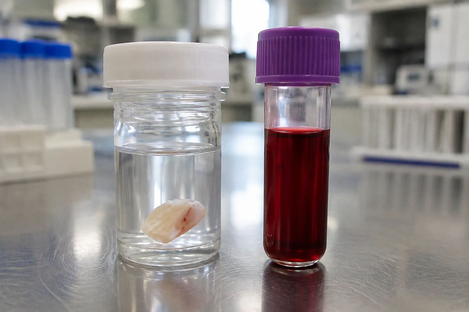
The throughput argument for liquid biopsy is straightforward. A blood draw requires no scheduling coordination with interventional radiology, no recovery time, no specimen transport logistics from procedure suite to pathology lab. A standard EDTA tube ships to the molecular lab the same day. From a supply chain perspective, the reduction in handling complexity alone improves operational efficiency.
But the clinical data sharpen the case considerably. A retrospective study evaluating 170 newly diagnosed NSCLC patients compared a liquid-first testing strategy against a tissue-first approach. The liquid-first cohort identified guideline-recommended biomarkers in 76.5% of patients. The tissue-first cohort managed 54.9%. That is a 21.6 percentage-point gap in first-pass biomarker capture — not a marginal improvement, but a structural inefficiency embedded in conventional workflows.
A 21.6 percentage-point gap in first-pass biomarker capture is not a marginal gain — it is a structural failure of tissue-first algorithms in advanced NSCLC.
For the lab director managing report turnaround benchmarks, the speed differential compounds the advantage. Genomic concordance between liquid biopsy NGS and tissue NGS for guideline-recommended biomarkers in NSCLC ranges from 94.8% to 100% in published cohorts. When the plasma assay agrees with tissue in nearly all cases, the operational case for front-loading the blood draw becomes difficult to contest.
| Parameter | Liquid-First Strategy | Tissue-First Strategy |
|---|---|---|
| Guideline-recommended biomarker capture (NSCLC) | 76.5% | 54.9% |
| Genomic concordance with tissue NGS | 94.8%–100% | Reference standard |
| Turnaround time advantage | 10–26.8 days faster | Baseline |
| Sample procurement complexity | Routine blood draw | Invasive biopsy procedure |
| Spatial heterogeneity capture | Systemic — reflects multiple tumor sites | Local — single lesion sampled |
| PD-L1 / IHC biomarker assessment | Not available via ctDNA | Required — tissue only |
| CHIP interference risk | Present — requires mitigation | Minimal for somatic variants |
Capturing Spatial Heterogeneity and Clonal Evolution
A tissue biopsy samples one site. One lesion. One region of one tumor. In a patient with stage IV disease — multiple metastatic sites, varying microenvironments, differential selective pressures — that single-core sample represents a snapshot, not a landscape. Spatial heterogeneity is not a theoretical concern. It is the reason certain patients with apparently driver-negative tissue results respond to targeted therapy when the alteration is detected in blood.
Plasma ctDNA is shed into the bloodstream by tumors across all anatomical sites. It integrates genomic information from primary and metastatic deposits simultaneously. For a lab building a precision oncology service line, this characteristic has direct operational value: it reduces the probability of a false-negative result driven by sampling bias, and it eliminates the need for repeat invasive procedures when initial tissue is insufficient for molecular analysis.
The implications extend into therapy monitoring and resistance profiling. Serial blood draws — logistically trivial compared to repeat biopsies — enable real-time tracking of clonal evolution under treatment pressure. For the hospital administrator evaluating capital allocation toward molecular diagnostics infrastructure, liquid biopsy capacity is not a luxury addition. It is a foundational capability for longitudinal patient management.
Liquid biopsy does not replace the scalpel. It replaces the wait — and the blind spots inherent in single-site sampling.
The CHIP Problem: When Blood Lies
No responsible analysis of plasma ctDNA testing omits the risk of clonal hematopoiesis of indeterminate potential. CHIP refers to age-related mutations arising in blood cells — not from the solid tumor — that are detectable in cell-free DNA. Without mitigation, these variants introduce false positives into the genomic profile, potentially misdirecting therapy selection toward targets the tumor does not actually harbor.
The compliance and liability implications are significant. A targeted therapy prescribed on the basis of a CHIP-driven false positive exposes the institution to clinical risk and potential regulatory scrutiny. Mitigation strategies exist — matched normal sequencing using a buffy coat sample, advanced bioinformatics pipelines that filter known CHIP-associated genes — but each introduces cost and workflow complexity.
For the lab director evaluating platform selection, CHIP filtering capability must sit alongside sensitivity and turnaround time as a non-negotiable criterion. The cheapest assay with the fastest TAT is a liability if it cannot reliably distinguish tumor-derived ctDNA from hematopoietic noise. Vendor evaluation frameworks must weight bioinformatics maturity as heavily as analytical performance specifications.
| CHIP Mitigation Strategy | Operational Impact |
|---|---|
| Matched normal sequencing (buffy coat) | Additional sample requirement; incremental sequencing cost |
| Bioinformatics filtering of known CHIP genes | Requires validated pipeline; ongoing curation burden |
| Tumor-informed assays (e.g., tumor-derived variants tracked in blood) | Higher upfront tissue requirement; superior specificity |
| Limiting reporting to guideline-tier alterations only | Reduces noise; may miss emerging targets |
Building the Diagnostic Algorithm: Actionable Framework
The optimal testing strategy is not liquid-only or tissue-only. It is algorithmic — designed to capture the maximum amount of actionable genomic information in the shortest time, without sacrificing histological confirmation or exposing the institution to compliance risk.
The framework, based on published evidence and guideline consensus, breaks down as follows:
1. Draw blood at first consultation. Plasma ctDNA NGS begins immediately. No scheduling delay, no procedural risk. The clock starts on day one.
2. Procure tissue in parallel. Histological diagnosis, tumor morphology, and PD-L1 immunohistochemistry require tissue. This step is mandatory and cannot be shortcut.
3. Use liquid biopsy results to initiate therapy when tissue is delayed or insufficient. If plasma NGS identifies an actionable alteration with high-confidence variant calling, treatment can begin while tissue processing continues — particularly critical in aggressive disease with rapid clinical deterioration.
4. Validate plasma findings against tissue when available. Concordance confirmation strengthens the clinical record and supports reimbursement documentation.
5. Implement CHIP-aware bioinformatics across all plasma-based reporting. No exceptions. The cost of a single misdirected therapy course dwarfs the investment in filtering infrastructure.
6. Establish serial ctDNA monitoring protocols for patients on targeted therapy. Resistance mutations detected in blood inform treatment pivots faster than repeat tissue biopsy.
This is not a theoretical exercise. The 2017 FDA tumor-agnostic approval of pembrolizumab for MSI-high tumors set the precedent for biomarker-driven, histology-agnostic prescribing. Every subsequent companion diagnostic approval tightens the operational requirement for rapid, reliable genomic profiling. Labs that cannot deliver will lose referral volume to those that can.
The Compliance and Margin Equation
For hospital administrators, the financial calculus is unambiguous. Every day of diagnostic delay in a patient eligible for targeted therapy represents unrealized treatment revenue, increased supportive care costs, and potential disease progression that drives more expensive downstream interventions. Liquid biopsy compresses that delay by 10 to 26.8 days.
The investment required — platform acquisition, bioinformatics staffing, CHIP filtering validation, paired sample logistics — is front-loaded but finite. The throughput gains are recurring. Labs that integrate a liquid-first algorithm into their standard-of-care pathway for advanced solid tumors position themselves to capture molecular testing volume that increasingly drives referral patterns and payer contracts.
Reimbursement structures continue to evolve, and the specifics of coverage for simultaneous paired liquid and tissue NGS testing vary across payers. That variability is a reason to track policy changes — not a reason to delay infrastructure buildout. The clinical guidelines are already ahead of the reimbursement curve. Labs operating on last decade's testing algorithm are leaving both margin and clinical outcomes on the table.
Every laboratory director overseeing molecular diagnostics for advanced solid tumors should be asking one question today: are we drawing blood at first consultation, or are we waiting for tissue? If the answer is the latter, the cost of that delay is already being paid — by patients and by the institution.