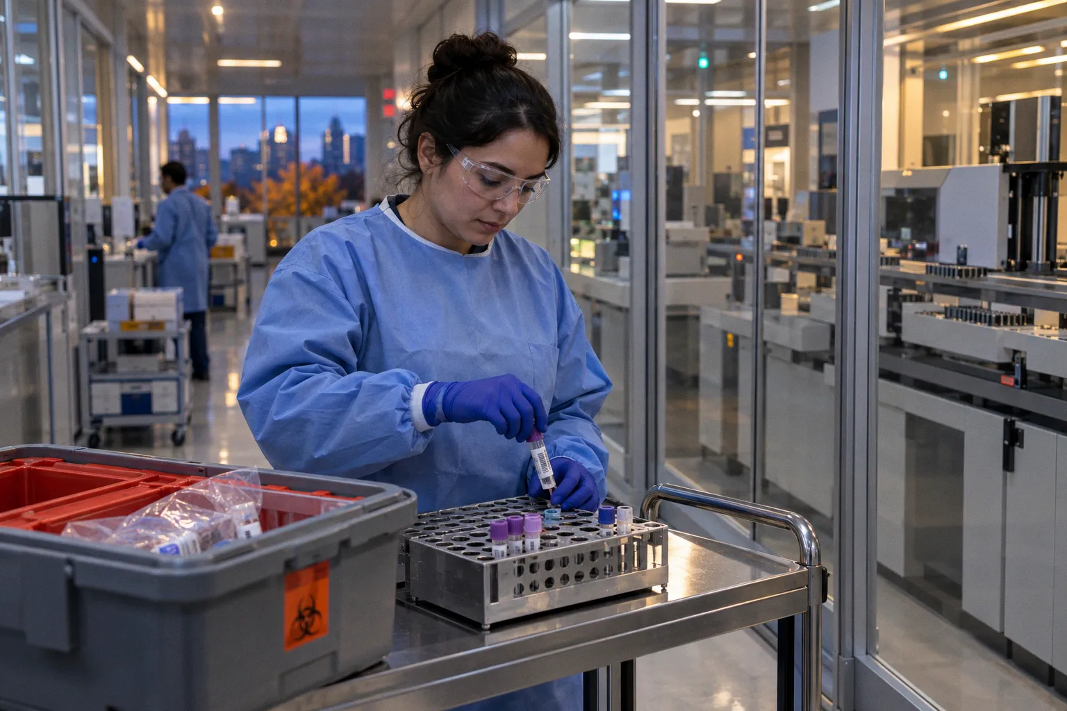
The clinical team knows there is a real process underway, but the laboratory has no organism to name, no colony to work up, and sometimes no clear target for a PCR assay. Antibiotics may already have altered the sample. The suspected pathogen may be unusual, present in very low numbers, or simply not included in the initial test menu.
This is where metagenomic next-generation sequencing for infectious disease diagnosis has become increasingly compelling. Instead of asking the laboratory to confirm one suspected organism, mNGS surveys nucleic acid in the specimen and looks broadly for microbial signatures. It is culture-independent and hypothesis-free, which can be transformative in complex infections. It is also expensive, vulnerable to host-DNA interference, and capable of finding material that is present but not actually causing disease.
That tension is the center of mNGS clinical utility in microbiology. The technology can widen the diagnostic window, but it does not remove the need for specimen judgment, contamination control, clinical context, or conventional microbiology. In many cases, it gives the bench a better question to pursue rather than a complete answer on its own.
Beyond culture: the shift to hypothesis-free diagnostics
Traditional microbiology is built around a sequence of reasonable bets. A clinician suspects bacterial pneumonia, a urinary infection, a bloodstream infection, or a wound pathogen. The laboratory selects media, incubation conditions, stains, biochemical tests, or molecular assays that fit the suspected clinical picture. When the hypothesis is good and the specimen is collected well, this approach remains powerful, relatively affordable, and directly connected to antimicrobial susceptibility testing.
The difficulty begins when the hypothesis is incomplete.
Culture may fail after antimicrobial exposure or when the organism is slow-growing, fastidious, intracellular, or difficult to recover from the specimen. Targeted PCR may be highly sensitive for the organisms it was designed to detect but cannot identify a pathogen outside that panel. A multiplex respiratory panel can answer a focused respiratory question; it cannot explain every inflammatory syndrome in a patient whose symptoms cross organ systems. The same limitation appears in gastrointestinal diagnostics, urogenital pathogen detection, and wound profiling: the panel is only as broad as its design.
mNGS changes the starting point. In a typical workflow, nucleic acid is extracted from a clinical specimen and sequenced without selecting a single pathogen in advance. Bioinformatic analysis then compares the resulting reads with reference databases and attempts to separate microbial sequences from human background and technical noise.
The phrase unbiased pathogen detection workflow is useful here, but it should not be mistaken for perfect neutrality. The method is broad in what it can seek. It is not free from bias. Extraction chemistry, library preparation, sequencing depth, database quality, organism biology, specimen type, and prior antimicrobial therapy all influence what can be detected.
A laboratory still has to make hands-on decisions before the sequencing run begins:
- Is the specimen from a normally sterile site, such as cerebrospinal fluid, blood, or a tissue compartment that should not contain microorganisms?
- Is the sample likely to carry abundant human DNA that will consume sequencing capacity?
- Was it collected from a non-sterile site where colonizing flora are expected?
- Was it transported and stored in a way that preserves the signal of interest?
- Is there enough material left for orthogonal confirmation and antimicrobial susceptibility testing?
Those questions may sound familiar to anyone who has worked with culture. They are familiar because the central discipline has not changed. The laboratory is still trying to connect a signal in a specimen with disease in a person.
mNGS expands the search space, but specimen quality and clinical reasoning still decide whether a detected organism means infection.
The practical advantage is speed in the right setting. Standard mNGS workflows typically produce a result in approximately 24 hours, with some workflows described in a range of roughly 7 to 24 hours. Traditional bacterial culture can require up to 5 days, particularly when recovery, identification, and follow-up work are all included. That difference matters when a patient is deteriorating and every additional incubation period carries clinical weight.
Speed, however, should be defined carefully. A sequencing result is not always the end of the diagnostic process. The laboratory may need to confirm the finding with targeted PCR, culture, Sanger sequencing, repeat testing, or another method. If treatment depends on susceptibility, mNGS alone generally does not replace antimicrobial susceptibility testing. A rapid organism call and a complete treatment profile are different laboratory products.
What the performance data show in complex infection
The strongest case for mNGS is not routine uncomplicated infection. It is the patient whose diagnostic pathway has become crowded with negative or discordant results.
In adult patients with fever of unknown origin, mNGS has demonstrated higher diagnostic sensitivity than conventional culture for infectious causes: 69.1% compared with 16.9%. That is a substantial difference in the ability to detect an infectious etiology. But the corresponding specificity was lower for mNGS, at 57.6% compared with 96.0% for conventional culture.
For a microbiologist, that combination is immediately recognizable. A highly sensitive method can find more true infections, but it can also find more signals that require interpretation. The sequencing result may reflect low-level contamination, colonization, residual nucleic acid, or a microorganism that is present without being responsible for the patient’s syndrome.
Tissue specimens show a similar pattern of promise. In patients with suspected infectious disease, tissue mNGS yielded a sensitivity of 72.7%, compared with 29.5% for conventional microbiological testing. Tissue can be a difficult matrix: organisms may be unevenly distributed, the available sample may be small, and prior treatment may have reduced culture recovery. A culture-negative tissue sample is not necessarily sterile tissue.
Still, these figures should not be read as a universal declaration that sequencing is superior for every specimen and every clinical question. Study populations, specimen handling, definitions of infection, sequencing platforms, and interpretation thresholds all shape observed performance. The numbers tell us where mNGS can add value. They do not eliminate the need for validation in the laboratory that intends to report it.
Where mNGS is most likely to add value
The clinical use case is strongest when several of the following conditions are present:
- The infection is serious, complex, or occurring in an immunocompromised patient.
- Conventional cultures are negative despite persistent clinical evidence of infection.
- Antibiotic, antifungal, or antiviral treatment began before specimen collection.
- The suspected organism is unusual, fastidious, or not covered by available targeted assays.
- The sample comes from a normally sterile site or from tissue with a strong clinicopathologic indication of infection.
- The result could change antimicrobial treatment, prompt source control, or redirect additional diagnostic work.
- Enough specimen remains for confirmation and, when needed, culture-based susceptibility testing.
This is different from ordering the broadest available test simply because the clinical picture is uncertain. Broad testing produces broad responsibility. The more organisms a method can detect, the more carefully the laboratory must explain what detection means.
mNGS versus targeted PCR: breadth is not the same as usefulness
The comparison between metagenomic sequencing and targeted PCR is often framed as old technology versus new technology. That framing is too blunt for the bench.
Targeted PCR is usually faster to validate for a defined organism or syndrome, easier to interpret, and more economical when the clinical suspicion is focused. If a patient has a presentation strongly suggestive of a pathogen covered by a validated assay, targeted PCR may provide the cleanest answer. It can also be more analytically sensitive for a particular organism than an untargeted approach, depending on the assay and specimen.
mNGS is most valuable when the differential diagnosis is wide, the initial workup is unrevealing, or the likely organism is not known in advance. It can detect bacterial, viral, fungal, and parasitic nucleic acid in the same broad survey, although performance varies by organism and specimen. The method may also reveal mixed infections that a single-target strategy would miss.
| Diagnostic approach | Main strength | Main limitation | Best-fit clinical setting |
|---|---|---|---|
| Culture | Provides viable organisms for identification and susceptibility testing | Slow or negative after treatment; may miss fastidious organisms | Suspected bacterial infection when viable recovery is realistic |
| Targeted PCR | Rapid, focused detection with straightforward interpretation | Cannot detect organisms outside the assay targets | Strong clinical suspicion for a defined pathogen or syndrome |
| Multiplex PCR panel | Efficient testing of several predefined pathogens | Fixed menu; positive result still requires clinical interpretation | Common respiratory, gastrointestinal, or other syndromic presentations |
| mNGS | Broad, culture-independent, hypothesis-free detection | High cost, host-DNA interference, contamination and colonization concerns | Complex, severe, unexplained, or diagnostically refractory infection |
| MALDI-TOF mass spectrometry | Rapid identification from a recovered isolate | Requires growth and an interpretable organism preparation | Routine identification of cultured bacteria and fungi |
The table also shows why mNGS should not be treated as a replacement for the rest of clinical microbiology. Culture produces an isolate that can often be tested further. Targeted PCR offers a precise answer when the target is well chosen. MALDI-TOF mass spectrometry remains highly useful once an organism has been recovered. These methods do different work at different points in the diagnostic chain.
In practice, the most effective model is often layered. A rapid targeted assay may answer the immediate question. Culture may provide an isolate for antimicrobial susceptibility testing. mNGS may be reserved for the cases where those routes are negative, incomplete, or contradicted by the patient’s clinical course.
That layered strategy also protects the laboratory from a common mistake: using a technically impressive result to solve the wrong clinical problem.
The workflow is more than a sequencing run
The phrase bioinformatics challenges in mNGS tends to draw attention to pipelines, databases, and read counts. Those are real challenges, but the workflow begins much earlier, at accessioning and specimen triage.
A weak specimen cannot be rescued reliably by deeper sequencing. In a low-biomass sample, a substantial proportion of the reads may come from the patient rather than the pathogen. Human host DNA interference can dilute microbial signal and consume sequencing capacity. In a respiratory, urinary, or open-wound specimen, the background may also include normal flora, colonizers, and environmental organisms.
That means a robust workflow needs controls and context at every stage:
1. Specimen selection and clinical information
The laboratory needs to know the body site, collection method, timing of antimicrobial exposure, immune status, and major clinical question. A result without specimen context is a list of sequences looking for a story.
2. Pre-analytical handling
Transport, storage, tissue homogenization, and nucleic-acid extraction can all influence yield. Small differences in how a specimen is divided may determine whether there is enough material left for confirmatory testing.
3. Negative and positive controls
Reagent contamination is a known concern in low-biomass sequencing. Controls help identify background signals introduced during extraction, library preparation, or handling.
4. Host-background assessment
The proportion of human reads affects how much useful microbial information can be recovered. A result that detects no pathogen may reflect true absence, insufficient microbial burden, or overwhelming host background.
5. Reference database and pipeline review
Taxonomic assignments depend on the quality and currency of reference databases. Closely related organisms may be difficult to distinguish, and a database can contain incomplete or unevenly annotated information.
6. Clinical and laboratory review
The final interpretation should be reviewed alongside microscopy, culture, targeted molecular tests, imaging, pathology, and the patient’s syndrome. Sequencing does not operate outside the diagnostic ecosystem.
The bench-level work remains visible in every one of these steps. Someone must assess the specimen, recognize a pre-analytical problem, question an unexpected organism, compare the result with the Gram stain or histopathology, and decide whether the signal deserves confirmation. Automation can make the process faster, but it does not make judgment unnecessary.
The economic and operational barrier
The financial case for mNGS is difficult because the technology is not simply a faster version of a conventional test. It requires sequencing capacity, extraction and library preparation, bioinformatics infrastructure, quality systems, trained personnel, and an interpretation process that can absorb ambiguous results.
The cost of mNGS testing is typically 10 to 20 times higher than that of most conventional infectious disease laboratory tests. That difference is substantial enough to shape utilization, reimbursement, and the design of clinical pathways. A hospital cannot treat every undifferentiated fever as a sequencing case without creating a considerable burden on both the laboratory and the health system.
The more useful question is not whether mNGS is expensive in isolation. It is whether the test changes management in a case where the alternatives have already consumed time, treatment, imaging, invasive procedures, or repeated sampling. That calculation will vary by setting and by healthcare system. Universal cost-effectiveness thresholds do not yet apply neatly across inpatient populations or international models of care.
Operational questions are just as important:
- Can the laboratory process specimens continuously or only in batched runs?
- Is there a defined pathway for urgent results?
- Who reviews potentially significant organisms before reporting?
- How are unexpected findings communicated to infectious disease physicians?
- Does the laboratory have access to culture, susceptibility testing, histology, and confirmatory PCR?
- How are negative results worded when the specimen had a high host-DNA burden?
- What happens when sequencing detects multiple organisms?
A 24-hour turnaround time is clinically meaningful only if the result reaches the care team in time to influence a decision. The clock includes accessioning, extraction, sequencing, analysis, review, and communication. It also includes the human time required to decide whether a finding is credible.
The value of mNGS is not measured by how many organisms it can name. It is measured by whether the right result arrives in time to change care.
Laboratories considering clinical validation of metagenomic panels need to define intended use with the same care applied to any other diagnostic service. A test designed for cerebrospinal fluid should not automatically be assumed to perform the same way in urine or an open wound. The specimen matrix, expected microbial burden, background flora, contamination risk, and clinical consequences of a false-positive interpretation all change from site to site.
The hardest interpretation: colonization, contamination, or infection?
This is where the promise of unbiased pathogen detection meets the practical limits of molecular diagnostics.
In a normally sterile body site, detection of microbial nucleic acid may carry considerable weight, particularly when it aligns with symptoms, imaging, inflammatory findings, and other laboratory results. Even there, contamination and residual nucleic acid cannot be ignored. The significance depends on the organism, abundance, specimen quality, timing, and clinical picture.
In a non-sterile outpatient site, the interpretive problem becomes more difficult. Urine, respiratory specimens, genital specimens, stool, and open wounds can contain organisms that are part of the resident microbiota, transient colonizers, or environmental carryover. A highly sensitive mNGS method may detect these signals readily. That can create the appearance of polymicrobial infection where the patient has colonization, contamination, or a different primary disease.
This matters directly for antimicrobial stewardship. If every detected organism is treated as a pathogen, broad sequencing can encourage broader therapy rather than more precise therapy. The test then succeeds analytically while failing clinically.
Interpretation should therefore be tied to a set of questions rather than a single read-count threshold. There is no universal threshold that cleanly separates pathogen from commensal across every specimen type. The laboratory and clinical team should consider:
- Whether the organism is biologically plausible for the syndrome and body site.
- Whether it is detected above the background seen in negative controls.
- Whether the result is reproducible in a second aliquot or by an orthogonal method.
- Whether the organism has been described in the relevant clinical context.
- Whether microscopy, culture, histology, imaging, or inflammatory markers support the finding.
- Whether the detected organism explains the patient’s illness better than alternative diagnoses.
- Whether acting on the result would improve therapy, source control, or infection prevention.
A sequence is evidence. It is not a diagnosis by itself.
The same principle applies to antimicrobial resistance detection. mNGS may identify genetic material associated with resistance, but the presence of a resistance determinant does not always establish its expression, its organism of origin, or the phenotype of the clinically relevant isolate. When viable organisms can be recovered, phenotypic AST and validated molecular resistance assays remain essential parts of the treatment decision.
Where this leaves the laboratory
mNGS is most persuasive when conventional approaches have been used thoughtfully and have not answered the question. It is particularly valuable in complex infection, immunocompromised patients, sterile-site and tissue specimens, and cases shaped by prior antimicrobial exposure. The performance data support that role: higher sensitivity in difficult clinical scenarios can uncover infections that culture misses.
But sensitivity without context can create a second diagnostic problem. A laboratory that adopts mNGS must invest not only in sequencing and computation but also in interpretation, communication, validation, and the quiet discipline of knowing when not to overcall a result.
I think of mNGS as an extension of microbiology rather than an escape from it. The method broadens the search, shortens some diagnostic timelines, and gives clinicians a way to investigate infections that resist conventional assumptions. Yet the decisive work still happens in the connection between specimen and patient: the quality of collection, the incubation history, the control result, the tissue finding, the culture plate, and the conversation around an unexpected organism.
For now, the best use of metagenomic sequencing is selective and deliberate. Use it when the clinical question is genuinely broad, when the consequences of a missed pathogen are serious, and when the laboratory has a plan for confirming and interpreting what it finds. In those circumstances, mNGS can do something genuinely valuable: not replace the culture bench, but give it a wider and more informed view of the infection in front of it.