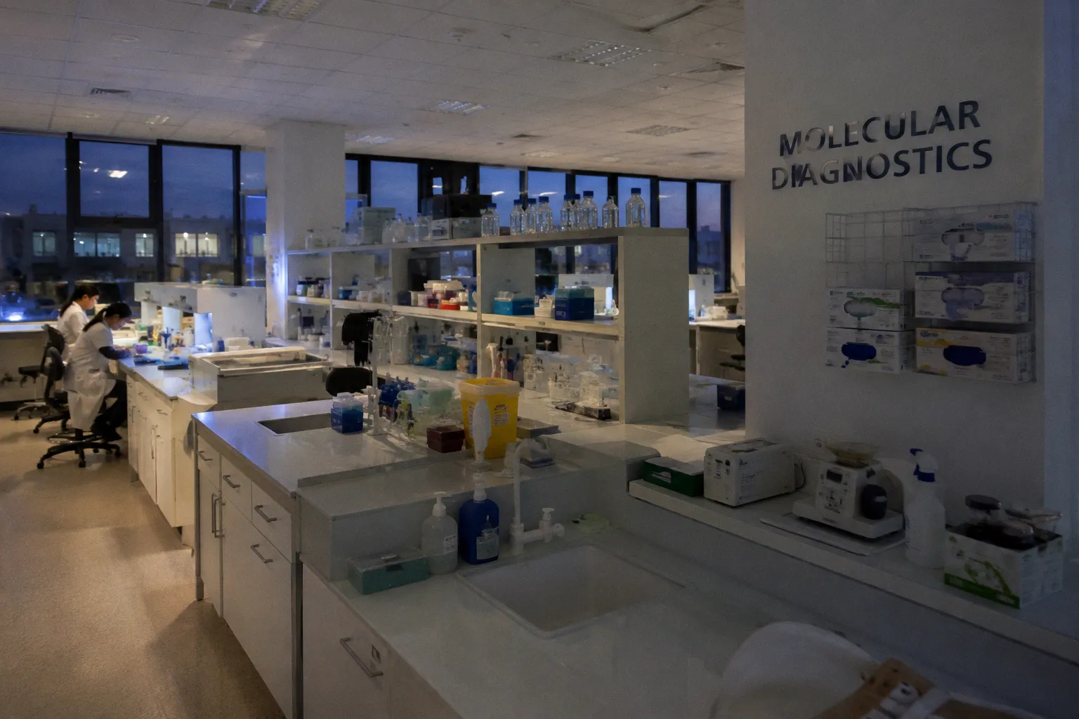
There is a call that comes across my desk with numbing regularity. A tumor-only NGS run finishes in the late afternoon, the bioinformatics pipeline produces its variant table, and somewhere in the file sits a single nucleotide change with a variant allele frequency hovering between two and five percent. The gene is often one we care about: a known driver, a resistance mutation, something the oncologist will ask about. The question that follows is the same every time: is this real tumor biology, or is this just what formalin does to damaged tissue?
I have learned to be skeptical of low-VAF calls in FFPE-derived DNA, and so has every molecular pathologist I have ever traded variant tables with. The reason sits in the chemistry of how we preserve tissue for the histology laboratory. Formalin — neutral buffered formaldehyde, the workhorse of every surgical pathology suite — is excellent at crosslinking proteins and preserving cellular architecture. It is much less gentle with the DNA inside those cells. Hydrolytic damage accumulates during fixation and storage, and one of its most consequential signatures is cytosine deamination.
That chemistry is responsible for a substantial share of the false-positive somatic variants encountered in FFPE sequencing. The artifacts are not random noise in the usual sense. They follow a recognizable pattern, appear disproportionately at low allele fractions, and can survive an otherwise competent variant-calling workflow if the laboratory does not account for their origin.
The Chemistry of Formalin-Induced Deamination: From Cytosine to Uracil
Every piece of DNA recovered from a slide or paraffin block carries a history. If the tissue spent too long in formalin, if fixation was uneven, or if the block was already highly degraded before extraction, cytosine bases may have undergone hydrolytic deamination. Water, time, and the chemical environment created during fixation all contribute to the process.
When cytosine loses its amino group, it becomes uracil. That distinction matters because uracil is still a readable base for many DNA polymerases. During PCR amplification, it can pair with adenine. After copying and subsequent amplification cycles, the original C:G base pair can therefore appear in sequencing data as a T:A substitution. In variant notation, this is the familiar C:G>T:A transition artifact — one of the characteristic formalin-induced sequencing artifacts in FFPE material.
The mechanism is worth following because it explains why the signal can look so convincing. Formaldehyde can react with primary amines on nucleotide bases and contribute to a damaged molecular environment in which base modifications and strand damage accumulate. Cytosine deamination itself is a hydrolytic reaction: the amino group is removed, leaving uracil in the DNA strand. The polymerase does not know whether that uracil was part of the original biological template or was created during fixation. It simply reads the base available to it and incorporates a complementary nucleotide.
The result is not necessarily a messy, low-quality read. A damaged molecule can have good mapping quality, sit neatly in a coding exon, and support a variant call at a clinically relevant locus. The variant caller sees aligned bases. The annotation pipeline sees a change in a gene. The report may then present a formalin-induced lesion as if it were a low-frequency tumor clone.
That is why the pattern matters more than any single read. A cluster of low-VAF C>T or G>A calls, especially when they occur near the ends of damaged molecules or are supported predominantly by one strand, should immediately raise the question of FFPE deamination artifacts in NGS testing.
A low-VAF mutation in FFPE-derived DNA is guilty until proven innocent — and the burden of proof usually falls on the laboratory, not the sequencer.
The intensity of the damage depends on several pre-analytical variables. Fixation time is the obvious one, but it is not the only one. Temperature, tissue thickness, fixative composition, tissue-to-formalin ratio, and the time between excision and fixation all shape the final DNA quality. Large resections are particularly difficult because formalin does not penetrate a thick specimen instantaneously. The outside of the tissue may be exposed to fixative long before the center.
The pH of the fixative also matters. Neutral buffered formalin is preferable to unbuffered formaldehyde because buffering limits some of the damaging chemistry, but it does not make the process chemically inert. A specimen that has been fixed for too long can still carry a considerable deamination burden even when the correct fixative was used.
Why the artifact is easy to mistake for a mutation
A real somatic mutation and a deamination artifact can share several superficial features:
- both may be present in only a small fraction of sequencing reads;
- both may occur in a clinically important gene;
- both may be supported by reads with apparently acceptable alignment quality;
- both may survive basic quality filters;
- both may produce a clean single-nucleotide substitution in the final variant table.
The difference lies in molecular history, not visual appearance. A true variant was present in the original tumor DNA. A deamination artifact was created during tissue handling or became visible only when damaged DNA was amplified. Conventional sequencing reads do not always retain enough information to distinguish those histories on their own.
Quantifying the Impact: Fixation Duration and VAF Overrepresentation
This is where the conversation with the surgical team usually starts. When I ask how long a resection sat in formalin before it was grossed, I am often met with a shrug. The answer matters more than many laboratories realize. Prolonged fixation is associated with a higher burden of DNA damage and an increased frequency of C:G>T:A substitutions compared with better-controlled fixation intervals. The exact effect depends on the specimen and assay, but the direction is consistent: more time in formalin generally means more opportunities for damage to accumulate.
The complication is that fixation duration is rarely uniform across a specimen. A mastectomy, colectomy, or large pelvic resection may remain in formalin for days because the tissue itself slows penetration. The surface can be adequately fixed while the center of a tumor remains under-fixed. Later, the outer regions may be over-fixed by the time the specimen is fully processed. DNA extracted from different blocks can therefore behave very differently even when the blocks came from the same operation and the same container of fixative.
This is one reason pathology review remains part of molecular interpretation. The molecular laboratory needs to know where the block came from, how much viable tumor it contains, whether necrosis is present, and whether the sampled region was likely to have experienced uneven fixation. A molecular result detached from specimen context is easier to overinterpret.
The problem becomes more consequential at low variant allele frequencies. A true heterozygous mutation present at a substantial fraction of tumor DNA is usually robust to a modest amount of background damage. The same is not true of a candidate variant at two percent VAF. At that level, the signal and the artifact burden may occupy the same analytical space.
This is particularly important when the assay is being used to search for subclonal resistance events, minimal residual disease, or other low-burden populations. The laboratory is deliberately looking below the level at which ordinary sequencing noise becomes negligible. In that setting, the question is not whether the sample contains damage. It is whether the workflow can distinguish a biological low-frequency signal from a chemical one.
The most useful pre-analytical information includes:
- the interval between excision and immersion in fixative;
- the duration of formalin fixation;
- the approximate thickness of the tissue during fixation;
- whether the block was prepared from a peripheral or central tumor region;
- the age and storage history of the paraffin block;
- the estimated tumor fraction and amount of necrosis;
- the DNA input and the extraction method used for the assay.
None of these variables proves that a call is false. They change the prior probability. A two-percent C>T variant from a heavily over-fixed, low-input block deserves more scrutiny than the same call from a well-controlled, high-tumor-content specimen.
| Pre-analytical variable | Likely effect on C:G>T:A artifacts | Practical consequence |
|---|---|---|
| Short, controlled fixation | Lower damage burden | Standard quality review may be adequate |
| Prolonged fixation | More deamination-related substitutions | Apply FFPE-aware filters and review strand support |
| Thick or poorly penetrated tissue | Spatially uneven DNA quality | Compare blocks and correlate with block location |
| Elevated temperature during handling | Can accelerate damaging reactions | Treat fixation records as part of molecular QC |
| Buffered formalin | Reduces some avoidable variability | Still does not eliminate deamination |
| Older or highly degraded blocks | Lower complexity and more damaged templates | Interpret low-VAF calls conservatively |
The practical mistake is to treat fixation as a historical detail rather than as an assay variable. It belongs beside DNA input, library complexity, coverage uniformity, and mapping quality in the interpretation workflow.
Enzymatic Mitigation: The Role of Uracil-DNA Glycosylase in Library Prep
This is where many laboratories reach for uracil-DNA glycosylase, usually abbreviated UDG or UNG depending on the laboratory’s terminology and protocol. UDG recognizes uracil bases in DNA and removes the damaged base, leaving an abasic site in the strand.
That abasic site is not a replacement nucleotide waiting to be chosen at random. It is a lesion in the DNA backbone that interferes with polymerase progression and can be cleaved or otherwise processed before amplification. When the workflow is designed correctly, the uracil-containing template is prevented from being copied as though it were a genuine cytosine-derived sequence. The objective is to remove the lesion from the amplification path, not to rewrite it with an arbitrary base.
This distinction is mechanistically important. UDG treatment does not repair the original DNA back to cytosine. It removes uracil and creates a substrate that must be handled by the library-preparation chemistry. Depending on the protocol, the abasic site may be cleaved or rendered non-amplifiable. In either case, the deaminated molecule is excluded or weakened before it can generate a misleading C:G>T:A signal during PCR.
UDG treatment is not a universal default in NGS library preparation. It is a workflow-specific intervention that must be deliberately included and validated for FFPE-derived material. General-purpose library kits designed for high-quality DNA from blood or fresh-frozen tissue may omit it because the substrate is not expected to be a major concern. FFPE-optimized workflows may include enzymatic treatment as part of the kit or specify it as an optional step that the laboratory must activate and validate.
That distinction matters. A laboratory cannot assume that a standard library kit neutralizes formalin-induced sequencing artifacts simply because the sample type is listed as compatible. Compatibility may refer to DNA yield, fragment size, or library construction success, not to suppression of low-VAF deamination artifacts.
What UDG can and cannot do
UDG is most useful when the artifact arises from uracil created by cytosine deamination. It is less useful when the false signal comes from a different lesion or from a technical error introduced later in the workflow. It also cannot recover information from a molecule that was lost during extraction, fragmented beyond practical use, or excluded because the uracil removal step made it non-amplifiable.
A sensible validation therefore looks beyond library yield. It should examine:
1. the frequency of C:G>T:A substitutions before and after treatment;
2. the effect on unique molecular complexity;
3. the behavior of low-VAF reference variants;
4. the effect on coverage uniformity across the target region;
5. the residual artifact pattern at CpG and non-CpG sites;
6. the performance of the complete workflow, including bioinformatic filtering.
The trade-off is real. Removing damaged molecules can reduce library complexity, particularly in specimens that already contain little intact DNA. A protocol that eliminates artifacts but leaves too few unique molecules for reliable calling has not solved the clinical problem. It has exchanged one failure mode for another.
For that reason, UDG concentration, incubation conditions, and timing should be treated as assay-validation parameters rather than universal settings. The right balance depends on the extraction method, DNA fragmentation profile, input amount, target design, and intended VAF range. A laboratory reporting variants below five percent VAF should know exactly how its UDG step changes both analytical sensitivity and artifact suppression.
UDG also does not replace specimen review or computational filtering. It reduces one important class of damage before amplification. It does not make an over-fixed block equivalent to a well-preserved sample.
The Methylation Blind Spot: Why CpG Sites Defy Standard UDG Repair
The most persistent limitation appears at methylated cytosines.
A fraction of cytosines in the mammalian genome carries a methyl group at the five-carbon position. These 5-methylcytosines are particularly common at CpG dinucleotides and have important roles in gene regulation and cellular identity. When 5-methylcytosine undergoes hydrolytic deamination, it becomes thymine, not uracil.
That difference is decisive. Thymine is a normal DNA base. Standard UDG does not recognize it as a damaged uracil and cannot remove it. A C>T change caused by 5-methylcytosine deamination therefore survives ordinary UDG treatment.
CpG sites are already mutation-prone in human genomes because spontaneous deamination of 5-methylcytosine occurs in living cells. FFPE processing can add another layer of damage on top of that biological background. In the final sequencing data, the laboratory may be unable to determine from the base substitution alone whether a C>T event reflects a genuine tumor mutation, a pre-existing biological change, or formalin-associated damage.
The enzyme catches the cytosines. Methylated cytosines become thymine, and thymine gives the enzyme nothing to recognize.
This is not a minor technical footnote. CpG sites occur in clinically important regions, including recurrently altered cancer genes. A low-frequency C>T call at a CpG context may be real, but its context should lower the laboratory’s willingness to accept a single-strand, low-complexity observation without additional support.
The appropriate response is not to discard all CpG variants. That would remove genuine biology along with artifacts. Instead, the assay should treat sequence context as one part of evidence. Position-specific artifact models, strand balance, read orientation, molecular family support, block-to-block concordance, and orthogonal confirmation can all contribute to the final interpretation.
Some laboratories raise reporting thresholds for especially problematic contexts. Others use empirically calibrated filters based on negative FFPE controls. Neither approach is perfect. The important point is that a single global VAF cutoff cannot account for the different chemical risks attached to different bases and sequence contexts.
Bioinformatic Resolution: Leveraging Unique Molecular Identifiers for Consensus Reads
The remaining work belongs to the bioinformatics pipeline. One of the most useful tools for low-frequency variant analysis is the unique molecular identifier, or UMI.
A UMI is a short molecular tag attached to an original DNA fragment before substantial amplification occurs. Reads carrying the same tag can be grouped as descendants of the same starting molecule. Instead of counting every sequencing read as independent evidence, the pipeline can build a consensus representation of the parent molecule.
That distinction is critical in FFPE material. If a damaged molecule is amplified many times, ordinary read counting can make one original lesion appear to be strong evidence. UMI-based analysis prevents that amplification history from being mistaken for independent molecular support. A family of twenty reads is not necessarily twenty independent observations. It may be one damaged template copied twenty times.
The most informative workflows go further and preserve strand information. A single-strand deamination event may be represented among descendants of one original strand without corresponding evidence on the complementary strand. Duplex or dual-strand consensus methods can require support from both strands of the original DNA molecule before accepting an ultra-low-frequency variant.
The concept is straightforward. The implementation is not. Reliable consensus calling requires sufficient family size, accurate UMI extraction, careful handling of sequencing errors, and enough unique molecules to represent the biological sample. Excessive PCR duplication does not create more evidence; it creates more copies of the same evidence. A library can therefore show impressive raw depth while containing too few independent molecules for confident low-VAF interpretation.
UMIs also increase the importance of pre-analytical quality. If the DNA was heavily fragmented before tagging, many molecules will not produce usable families. If the tumor fraction is low, the number of unique tumor-derived molecules may be inadequate even when total read depth is high. If the assay reports a variant near its limit of detection, the laboratory should evaluate unique molecule counts and consensus support, not only the conventional depth displayed by the variant caller.
What a convincing low-VAF call should show
There is no single feature that proves a low-frequency FFPE variant is genuine. Confidence comes from several independent observations aligning:
- support from multiple unique molecular families rather than many duplicate reads;
- balanced representation across read directions where the assay permits that assessment;
- evidence from both original DNA strands in a duplex workflow;
- absence of a strong end-of-read or damage-associated pattern;
- a sequence context that is not disproportionately represented among the sample’s other artifacts;
- adequate tumor fraction and sufficient unique molecule input;
- consistency across technical replicates, blocks, or orthogonal assays when clinically necessary.
The absence of one feature does not automatically invalidate a call. A targeted panel may have uneven strand design. A very small specimen may not support an independent replicate. A true mutation near a read end may be unavoidable because of the amplicon structure. Interpretation has to be tied to the validated design of the assay rather than to a generic checklist imported from another platform.
UMIs do not replace UDG. They complement it. UDG removes a major class of uracil-containing templates before amplification. UMIs limit the ability of one damaged molecule to masquerade as many independent observations and can expose strand-specific damage. Neither tool corrects every FFPE problem. Together, however, they address different stages of the same analytical threat.
Bringing the chemistry back to the report
The final decision should not be made from the variant table alone. A molecular pathologist reviewing a suspicious FFPE call needs to connect the chemistry to the specimen and to the assay.
A low-VAF C>T or G>A variant from a severely over-fixed block, with weak unique molecule support and strong strand imbalance, should not be reported with the same confidence as a variant supported by independent duplex families in a well-controlled specimen. At the other end of the spectrum, a variant that survives UDG treatment, appears across independent molecules and both strands, fits the tumor’s biology, and is reproduced by an orthogonal method may be entirely credible even if the initial specimen was not ideal.
The wording of the report should reflect that distinction. When the evidence is limited, the laboratory can describe the result as potentially affected by FFPE-associated damage and recommend confirmation rather than presenting an uncertain low-VAF call as established tumor biology. When the evidence is strong, the report can explain why the laboratory considers the finding reliable. The goal is not to make every difficult variant disappear. It is to make the uncertainty visible before it reaches a treatment decision.
This is also why fixation records belong in the molecular diagnostic conversation. A pathology laboratory that tracks fixation duration, tissue dimensions, block selection, DNA input, unique molecule counts, and artifact signatures is not collecting administrative trivia. It is building the evidence needed to interpret the result.
FFPE deamination artifacts in NGS testing are not a nuisance that can be solved by one universal filter. They are the predictable consequence of exposing biological material to a chemical preservation process and then asking damaged molecules to support low-frequency clinical conclusions. UDG can remove uracil-derived lesions. It cannot repair methylated-cytosine deamination. UMIs can reveal amplification history and strengthen consensus calling. They cannot create independent molecules that were lost before library preparation. Bioinformatics can model the residual noise. It cannot recover information that the specimen never preserved.
The reliable answer comes from treating the entire workflow as one system: tissue handling, fixation, extraction, library preparation, enzymatic mitigation, molecular barcoding, consensus analysis, and pathology review. A low-VAF mutation is not real because a sequencer printed it in a table. It becomes convincing when the chemistry, the molecules, the specimen context, and the clinical interpretation all point in the same direction.