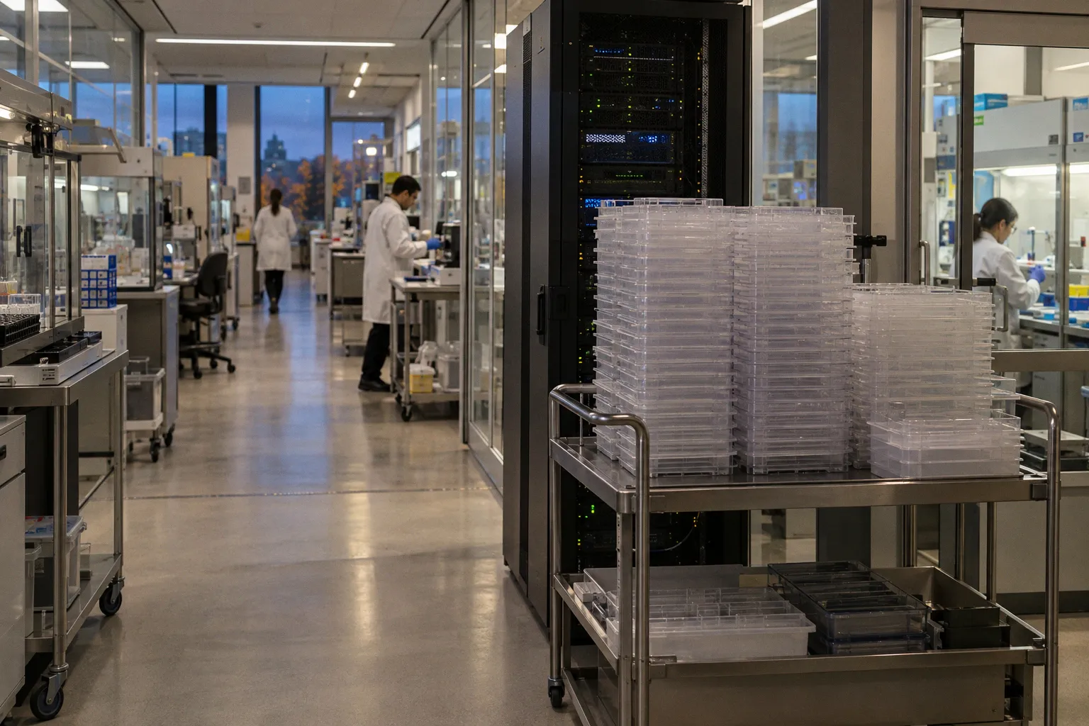
Large specimens — prostatectomies, wide breast excisions, multi-tissue blocks — routinely exceed 8 GB and can push toward 12 GB. Multiply that by tens of thousands of slides a year and the archive problem starts to look less like a laboratory upgrade and more like an infrastructure program.
The digital transformation of pathology is real, but the bytes are louder than the brochures.
A lab digitizing 10,000 slides a year may generate approximately 50 TB of new image data annually. High-throughput operations — academic medical centers, national reference laboratories, and hospital networks using whole-slide imaging as a routine part of practice — can move beyond 500 TB a year. That growth does not stop when the scanner is paid off. Storage becomes a recurring operational expense, and the cost is shaped as much by retrieval patterns, retention rules, and network design as by the scanner itself.
Vendors will often frame the cloud as the answer. Sometimes it is part of the answer. It is not the architecture. The practical solution is usually a combination of tiered storage, deliberate compression, controlled file formats, and a retention policy that has been written, approved, and tested rather than left to grow accidentally.
The Physics of Gigapixel Data: Understanding WSI File Architecture
A whole slide image is not simply a very large photograph. It is a multi-resolution object designed to let a pathologist pan and zoom without loading the entire slide into memory. During acquisition, the scanner divides the tissue into tiles and creates several representations of the same image at different magnification levels. The viewer then requests the tiles it needs for the current field of view and zoom level.
That architecture is what makes diagnostic viewing responsive. It is also why a WSI file is substantially more demanding than a conventional microscope photograph.
Two resolutions are common in routine digital pathology workflows. At 40×, the optical sampling is approximately 0.25 micrometers per pixel; at 20×, it is roughly 0.50 micrometers per pixel. The difference is not cosmetic. Moving from 20× to 40× increases the number of pixels that represent the same tissue area and, in turn, increases the storage and processing burden.
The storage impact depends on more than nominal magnification. Tissue area, scanner settings, focus strategy, z-stacking, barcode and metadata payloads, compression format, and the number of pyramid levels all influence the final file. A small biopsy scanned at 40× and a large resection scanned at the same nominal magnification are not equivalent storage objects.
Display infrastructure matters as well. A 2K display may be adequate for some viewing tasks, while 4K is increasingly expected for stations used in primary diagnosis. But the monitor is only one part of the chain. A high-resolution display does not compensate for a slow image server, an overloaded storage network, or a viewer that has to reconstruct pyramid levels on demand.
A whole slide image is not a document. It is a gigapixel object that needs the same architectural discipline as a sequencing run or an imaging study.
Why the pyramid consumes so much space
The full-resolution layer is only the most obvious part of the file. To support navigation, the WSI also stores lower-resolution versions, usually arranged as a pyramid. Each level covers the tissue at a different scale, and the viewer selects the most appropriate level before requesting individual tiles.
Those lower-resolution levels are not free. Their combined footprint is smaller than the base layer, but it is still substantial. A scan that appears to require a certain amount of storage based only on its full-resolution image will need additional capacity for the pyramid, metadata, thumbnails, labels, and sometimes multiple focus or z-stack planes.
The trade-off is straightforward:
- Precomputed pyramid levels increase the stored footprint but make navigation faster and more predictable.
- Fewer stored levels reduce capacity requirements but shift more work to the viewer or server.
- On-the-fly downsampling can be reasonable for selected workflows, but it becomes a problem when many users are opening large cases simultaneously.
- Z-stacking and extended focus acquisition can multiply the data burden because the system stores more than one focal plane for the same tissue area.
This is why a lab should not ask only how large the scanner’s “average file” is. It should ask what the average file contains, how the scanner creates it, and which parts of the image are required for diagnosis, consultation, artificial intelligence, or research.
Quantifying the Digital Burden: From 50TB to 500TB Annual Growth
The arithmetic is not difficult. The assumptions are.
A department digitizing 10,000 slides per year and producing an average of approximately 5 GB of compressed data per slide will generate about 50 TB of new storage demand annually. That figure does not include redundant copies, backups, disaster-recovery replicas, temporary processing files, or additional derivatives created by image-analysis systems.
At 100,000 slides per year, the same average would imply roughly 500 TB of new image data. Even if the laboratory’s actual average is lower, the conclusion does not change: the archive can grow by hundreds of terabytes before the organization has fully adapted its operating model.
The average, however, can be a misleading planning tool. WSI data is driven by tissue area and acquisition protocol more than by the slide count alone. A small biopsy core may produce a file near the lower end of the usual range. A large resection with multiple tissue fragments, extended focus, or z-stacks can reach 8 to 12 GB or more after compression. A laboratory that scans a high proportion of large specimens will therefore outgrow a capacity plan based on a generic per-slide estimate.
Case mix should be part of the storage model from the beginning. At minimum, the planning exercise should separate:
- Small biopsies and cores.
- Routine resections.
- Large excisions and multi-part specimens.
- Slides acquired at 20× versus 40×.
- Standard scans versus extended-focus or z-stack acquisitions.
- Primary diagnostic images versus research, teaching, or quality-control scans.
- Original files versus derivatives created for AI or quantitative analysis.
The distinction between primary images and derivatives is particularly important. A laboratory may store the original WSI, a compressed copy for routine access, thumbnails, tissue masks, annotations, algorithm outputs, and exported images for multidisciplinary review. If those objects are counted as “one slide” in a procurement conversation, the resulting capacity estimate will be optimistic by design.
Capacity planning is a workflow problem
Storage calculations should include the full lifecycle of the image:
1. Acquisition: the scanner writes the original image and associated metadata.
2. Quality control: the laboratory may retain failed scans, rescans, or images awaiting review.
3. Clinical use: the image is accessed repeatedly during sign-out, consultation, and case discussion.
4. Analysis: algorithms may create annotations, heat maps, probability maps, or derived measurements.
5. Backup and recovery: additional copies are required if the image is part of a clinically important record.
6. Archiving: closed cases move to a lower-cost tier but remain discoverable and retrievable.
7. Deletion or disposition: the organization applies its retention policy, if the image is eligible for removal.
A capacity plan that accounts only for the first step is not a storage plan. It is a scanner-output estimate.
Storage is not a one-time line item. It is a recurring operating expense shaped by acquisition volume, access patterns, redundancy, and the decisions made after sign-out.
Cloud storage costs for digital pathology can look attractive when quoted as a monthly price per terabyte. That number is only one component. Egress fees, replication, request charges, lifecycle transitions, network connectivity, retrieval time, and the cost of keeping data in a particular region can materially change the total. On-premises infrastructure has its own less visible costs: power, cooling, hardware refreshes, support contracts, backup systems, and the staff needed to operate them.
The useful comparison is not cloud versus on-premises in the abstract. It is the cost of a particular architecture for a particular workload.
Tiered Storage Strategies: Balancing Hot Access and Cold Archiving
No laboratory needs its entire WSI archive on high-performance flash. Few laboratories can treat the entire archive as genuinely cold either. The practical model is a tiered architecture in which storage performance follows clinical relevance and access frequency.
Hot storage is designed for active work. It may be backed by NVMe drives, high-performance solid-state storage, or a fast storage-area network. The tier should hold cases that are being signed out, recently reviewed cases, active consultations, current immunohistochemistry workups, and images likely to be opened repeatedly by several users.
Warm storage is useful for material that is not in the immediate diagnostic queue but still needs reasonably quick access. This can include recently closed cases, ongoing consultations, quality-assurance sets, or cases connected to a current clinical program. The exact definition will differ by laboratory, but the principle is consistent: not every archive object needs the same latency, and not every retrieval should trigger a lengthy restore process.
Cold storage is intended for infrequently accessed material. It may use lower-cost object storage, a cloud archive tier, or an on-premises tape library. Closed cases, historical consultation material, teaching sets, and completed research cohorts may belong here, provided that the laboratory can still locate and retrieve them when the policy requires it.
| Parameter | Hot storage | Warm storage | Cold storage |
|---|---|---|---|
| Typical media | NVMe SSD, fast SAN, high-performance object storage | Standard SSD, high-capacity disk, accessible object storage | Archive object storage or tape |
| Typical access | Frequent and interactive | Occasional, but still operational | Rare or policy-driven |
| Retrieval expectation | Near-immediate | Usually prompt | Seconds to minutes, or longer depending on the medium |
| Typical content | Active diagnostic cases, current consults | Recently closed cases, ongoing projects | Long-term archive, teaching sets, completed cohorts |
| Main design priority | Low latency and concurrency | Balance of cost and availability | Capacity, durability, and retention |
The tier boundary should not be determined by an arbitrary storage date alone. It should reflect workflow. A case that is technically closed may still be needed for a tumor board, a referral review, a discrepancy investigation, or a follow-up comparison. Conversely, some images become unlikely to be opened immediately after sign-out.
The migration trigger can be based on case closure, sign-out status, the end of a consultation window, or an institutional policy. What matters is that the transition is predictable and reversible. A pathologist should not discover that a clinically relevant image has been moved to a slow archive only when the image is urgently needed.
The network is part of the storage tier
A cold tier is not genuinely cheap if every retrieval requires manual intervention or creates a clinical delay. Moving a 5 GB slide from an archive bucket in another region may take minutes on a constrained connection. Retrieving several large slides for a consultation can create a much larger problem than the individual file size suggests.
The archive therefore has to be evaluated as a system:
- How long does it take to locate a case?
- How long does it take to restore the image?
- Can several users retrieve slides at the same time?
- Does the viewer support progressive loading?
- What happens when the network connection is interrupted?
- Is the restored copy automatically removed, or does it remain on the hot tier indefinitely?
- Are the audit trail and chain of custody preserved during migration?
A laboratory that expects fast random access to every case from the last decade has, in practical terms, made much of its archive “hot.” That may be an entirely reasonable decision, but it should be budgeted honestly.
Optimizing File Footprints: Compression and Pyramid Structures
Uncompressed WSI files can range from approximately 15 GB to well above 100 GB per slide, depending on tissue area, resolution, and acquisition mode. Those are not usually the files retained for everyday use. Compression is what makes routine digital pathology storage feasible.
JPEG and JPEG2000 are common components of WSI workflows. DICOM can provide a vendor-neutral framework for image exchange and associated metadata, while scanner manufacturers may also use proprietary pyramidal formats and codecs. The format question is not merely technical. It affects interoperability, migration, viewer performance, algorithm compatibility, and the laboratory’s ability to retrieve images after a vendor change.
Lossless and lossy compression
Lossless compression preserves the original pixel values after decompression. It is the safer choice for workflows that depend on exact image data, including certain quantitative measurements, algorithm development, validation datasets, and applications where small intensity differences may matter.
Lossy compression discards some information to achieve a smaller file. With appropriate settings, the visual difference may be difficult to detect during routine review, and the reduction in storage demand can be meaningful. But “not visible to the human eye” is not the same as “irrelevant to an algorithm.” A model may respond to artifacts, edge changes, texture shifts, or altered color relationships that are not obvious during manual inspection.
That does not make lossy compression unusable. It means that the compression regime should be treated as part of the image-processing environment. If a model was developed and validated on images produced with one codec or quality setting, changing the scanner configuration or archive pipeline can alter the input distribution. The change should be evaluated before it becomes part of a clinical workflow.
Compression decisions should therefore be linked to use case:
- Routine diagnostic review: a validated lossy setting may be appropriate if image quality remains acceptable for the intended task.
- AI inference: use the compression conditions covered by the model’s validation, or assess performance after a change.
- AI training: keep the data-generation process documented and consistent; retain representative original images where feasible.
- Quantitative pathology: prefer a format and compression method compatible with the measurements being performed.
- Long-term archiving: prioritize documented, supportable formats and preserve enough metadata to reconstruct how the image was created.
The key operational mistake is allowing settings to drift. A scanner is replaced, a software update changes the default quality factor, or an archive migration recompresses images to reduce capacity. Nobody records the change because the images still look normal in the viewer. Months later, an algorithm behaves differently or a quantitative pipeline produces results that are difficult to compare with older cases.
The pyramid is also a design decision
The number of pyramid levels influences both storage and responsiveness. A system that stores many precomputed levels generally offers predictable navigation, especially when pathologists move rapidly between overview and high-power views. A system that stores fewer levels may save space but require additional computation during viewing.
There is no universal optimum. The right balance depends on:
- The scanner’s native output.
- The viewer’s ability to generate or cache missing levels.
- The number of simultaneous users.
- The available CPU, GPU, and network capacity.
- Whether images are viewed locally or across a wide-area connection.
- Whether the same files are used for diagnosis, AI, education, and research.
Before procurement, the laboratory should test representative slides rather than a small biopsy selected for convenience. The test set should include large resections, high-cellularity tissue, slides with substantial background, and any acquisition modes the service expects to use. Storage requirements based on a single “typical” slide are rarely typical enough.
A short list of decisions is worth making before large-scale scanning begins:
1. Define which workflows require lossless data and which can use validated lossy compression.
2. Record the codec, quality settings, pyramid structure, scanner software version, and acquisition parameters in the standard operating procedure.
3. Test the same representative cases through the scanner, archive, viewer, and retrieval process.
4. Confirm that the chosen format can be exported or migrated without losing essential metadata.
5. Include pyramid overhead, derivatives, backups, and disaster-recovery copies in the capacity model.
6. Treat any change to compression parameters as a validation event for affected clinical and AI workflows.
7. Monitor file sizes over time so that a gradual change in acquisition settings does not become a surprise capacity crisis.
The goal is not to achieve the smallest possible file. It is to achieve a file that is small enough to manage, rich enough for its intended use, and documented well enough to remain trustworthy.
Navigating Retention Policies in a Post-Glass Slide Era
The legal and operational status of a digital slide is not always identical to that of the glass slide from which it was created. In many jurisdictions, retention rules were written around physical specimens and specify how long glass slides, blocks, or other pathology materials should be retained. Those rules may not describe the digital surrogate with the same precision.
That does not mean a digital archive can be deleted casually. It means the laboratory needs to define its own policy rather than assuming that the glass-slide rule automatically answers every question about WSI.
A retention policy should distinguish between at least three considerations:
- Clinical utility: whether the image may be needed for future comparison, consultation, review, or quality assurance.
- Institutional obligations: how the organization handles records, investigations, education, and research data.
- Technical and legal defensibility: whether the archive can demonstrate what was stored, when it was created, how it was modified, and who accessed it.
Different categories of images may reasonably have different retention paths. A teaching archive can be preserved for a long time because it remains actively useful. A research cohort may have a defined lifecycle tied to the study and its governance. A clinical image may be retained according to institutional policy, the role it plays in the medical record, and the likelihood that it will be needed for future care.
The policy should also address the relationship between the digital image and the physical material. If the glass slide is retained but the WSI is deleted, the laboratory may still be able to recover the information by rescanning — assuming the physical material remains intact, discoverable, and available. If the glass slide is no longer available, deleting the digital surrogate may be irreversible. That distinction belongs in the risk assessment.
Long-term archiving of histopathology images
Long-term archiving is not just a question of putting files in a low-cost bucket. The archive must remain intelligible. A WSI without reliable case identifiers, specimen relationships, acquisition metadata, and a functioning retrieval path is technically present but operationally lost.
A durable archive should account for:
- Stable identifiers linking the image to the case and specimen.
- Metadata describing scanner, resolution, magnification, and acquisition mode.
- Checksums or another method for detecting corruption.
- Multiple copies or an appropriate durability strategy.
- Periodic restoration tests rather than a one-time backup declaration.
- A documented process for migrating data when storage media or vendors change.
- Access controls and audit trails appropriate to clinical information.
- Clear rules for deletion, legal holds, research governance, and exceptions.
The restoration test is where many archives become theoretical. A backup that has never been restored is an assumption, not evidence. The laboratory should periodically select archived slides, retrieve them through the normal process, open them in the intended viewer, and confirm that the metadata and associated case context remain usable.
Retention decisions should be reviewed with compliance, legal, pathology leadership, information security, and IT. The point is not to retain everything forever. Indefinite retention is not automatically safer; it creates cost, security exposure, and governance obligations. The point is to make deletion and preservation deliberate decisions rather than accidental consequences of a storage tier reaching capacity.
The practical conclusion
The storage problem created by whole slide imaging is solvable, but not by buying more disk and not by signing a cloud contract in isolation. It is an architectural problem.
A workable design follows access patterns, separates active cases from long-term archives, uses compression according to the job the image has to perform, and treats pyramid construction as part of the file-footprint decision. It also recognizes that a slide may generate more than one object: the original image, derivatives, annotations, algorithm outputs, backups, and recovery copies all belong in the model.
The technical choices should be tied to clinical reality. A pathologist reviewing an active case needs predictable interactive performance. An AI team needs a documented and stable image-generation process. A compliance team needs a retention rule that can be explained. An IT team needs a capacity forecast that includes replicas and retrieval, not just the output reported by the scanner.
The difference between a manageable archive and a costly one is usually not a single codec, storage vendor, or scanner model. It is whether the laboratory has made its assumptions visible. Once the organization knows which images must be immediately available, which can move to cold storage, which workflows require lossless data, and how long each category should remain accessible, the terabytes become an engineering problem.
Without those decisions, the archive grows by default: more images, more copies, more derivatives, and less certainty about what can be deleted. With them, whole slide imaging becomes what it should have been from the beginning — not an uncontrolled accumulation of enormous files, but a governed clinical information system.